ReadiUse™ Rapid Luminometric ATP Assay Kit
Ordering information
Price | |
Catalog Number | |
Unit Size | |
Quantity |
Additional ordering information
Telephone | 1-800-990-8053 |
Fax | 1-800-609-2943 |
sales@aatbio.com | |
Quotation | Request |
International | See distributors |
Shipping | Standard overnight for United States, inquire for international |
Storage, safety and handling
H-phrase | H303, H313, H333 |
Hazard symbol | XN |
Intended use | Research Use Only (RUO) |
R-phrase | R20, R21, R22 |
Storage | Freeze (< -15 °C); Minimize light exposure |
UNSPSC | 12352200 |
Related products
Overview | ![]() ![]() |
See also: Cell Proliferation Assays
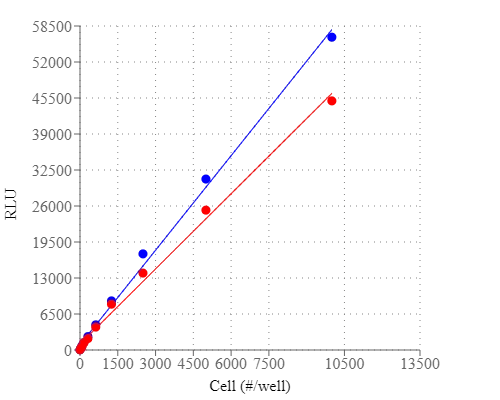
Adenosine triphosphate (ATP) plays a fundamental role in cellular energetics, metabolic regulation and cellular signaling. The quantitation of ATP can be used for a variety of biological applications. Because ATP is the energy source for almost all living organisms that rapidly degrades in the absence of viable organisms, its existence can be used to identify the presence of viable organisms. The measurement of ATP has been used for cell cytotoxicity, detection of bacteria on surfaces, quantification of bacteria in water, somatic cells in culture and food quality. The use of firefly bioluminescence to measure ATP was first proposed by McElroy when he discovered that ATP was essential for light production. Firefly luciferase is a monomeric 61 kD enzyme that catalyzes a two-step oxidation of luciferin, which yields light at 560 nm. The first step involves the activation of the protein by ATP to produce a reactive mixed anhydride intermediate. In the second step, the active intermediate reacts with oxygen to create a transient dioxetane, which quickly breaks down to the oxidized product oxyluciferin and carbon dioxide along with a burst of light. When ATP is the limiting component, the intensity of light is proportional to the concentration of ATP. Thus the measurement of the light intensity can be used for quantifying ATP using a luminometer. AAT Bioquest's ReadiUse™ Rapid Luminometric ATP Assay Kit (DTT free) comes with all the essential components in a ready-to-use format. It provides a fast, simple and homogeneous luminescence assay for monitoring cell proliferation and cytotoxicity in mammalian cells. This assay is based on the detection of ATP using firefly luciferase to catalyze the release of light by ATP and luciferin. It can be performed in a convenient 96-well or 384-well microtiter-plate format on a chemiluminescent microplate reader. The assay is extremely sensitive and can detect 50 cells/well. It has stable luminescent signal with half-life more than 2 hours. This ReadiUse™ Rapid Luminometric ATP Assay Kit does not use DTT, which eliminating the unpleasant odor.
Platform
Luminescence microplate reader
Recommended plate | Solid white |
Example protocol
AT A GLANCE
Protocol Summary
- Prepare cells (samples) with test compounds (100 µL/96-well plate or 25 µL/384-well plate)
- Add equal volume of ready-to-use ReadiUse™ Rapid Luminometric ATP Assay Reagent (100 µL/96-well plate or 25 µL/384-well plate)
- Incubate at room temperature for 10 - 20 minutes
- Monitor the luminescence intensity
Important notes
To achieve the best results, it’s strongly recommended to use the white plates. Thaw all the kits components at room temperature before starting the experiment.
PREPARATION OF STANDARD SOLUTION
ATP standard
For convenience, use the Serial Dilution Planner: https://www.aatbio.com/tools/serial-dilution/21601
Optional, ATP is not provided.
Make 1 mM ATP stock solution with ddH2O or appropriate buffer, then perform serial dilution to achieve ATP concentrations ranging from 10 pM to 10 µM.
For guidelines on cell sample preparation, please visit
https://www.aatbio.com/resources/guides/cell-sample-preparation.html
SAMPLE EXPERIMENTAL PROTOCOL
Table 1. Layout of ATP Standards and test samples in a white 96-well microplate. SD=ATP Standard, BL=Blank Control, TS=Test Sample
BL | BL | TS | TS |
SD1 | SD1 | ... | ... |
SD2 | SD2 | ... | ... |
SD3 | SD3 | ||
SD4 | SD4 | ||
SD5 | SD5 | ||
SD6 | SD6 | ||
SD7 | SD7 |
Table 2. Reagent composition for each well
Well | Volume | Reagent |
SD1-SD7 | 100 µL | Serial Dilution of ATP (e.g. 10 pM to 10 µM) |
BL | 100 µL | ATP Assay Buffer (Component A) |
TS | 100 µL | Sample |
- Treat cells (or samples) with test compounds by adding 10 µL of 10X compounds for a 96-well plate or 5 µL of 5X compounds for a 384-well plate in desired compound buffer. For blank wells (medium without the cells), add the corresponding amount of compound buffer.
- Incubate the cell plate in a 37 oC, 5% CO2 incubator for the desired period of time, such as 24, 48 or 96 hours.
- Add 100 µL/well (96-well plate) or 25 µL/well (384-well plate) of ReadiUse™ Rapid Luminometric ATP Assay Reagent.
- Incubate at room temperature for 10 - 20 minutes. Note: Aliquot and store the unused ReadiUse™ Rapid Luminometric ATP Assay Reagent at -20 oC, and avoid repeated freeze/thaw cycles.
- Monitor the luminescence intensity with a standard luminometer.
Images

Figure 1. Cell number correlates with the luminescent signal. Different Jurkat cell number (2-fold dilution) was measured using the ReadiUse™ Rapid Luminometric ATP Assay Kit in a 96-well white plate using a ClarioStar plate reader (BMG Labtech). The kit can detect as low as 50 cells. There is a linear relationship (r2 >0.99) between the luminescent signal and cell number after 15 minutes or 2 hours incubation. The half-life of luminescent signal is more than 2 hours.

Figure 2. Cell number correlates with the luminescent signal. Different Jurkat cell number (2-fold dilution) was measured using the ReadiUse™ Rapid Luminometric ATP Assay Kit in a 96-well white plate using a ClarioStar plate reader (BMG Labtech). The kit can detect as low as 50 cells. There is a linear relationship (r2 >0.99) between the luminescent signal and cell number after 15 minutes or 2 hours incubation. The half-life of luminescent signal is more than 2 hours.
Citations
View all 12 citations: Citation Explorer
Guidelines for cell viability assays
Authors: Kamiloglu, Senem and Sari, Gulce and Ozdal, Tugba and Capanoglu, Esra
Journal: Food Frontiers (2020): 332--349
Authors: Kamiloglu, Senem and Sari, Gulce and Ozdal, Tugba and Capanoglu, Esra
Journal: Food Frontiers (2020): 332--349
High throughput cell-based assay for identification of glycolate oxidase inhibitors as a potential treatment for Primary Hyperoxaluria Type 1
Authors: Wang, Mengqiao and Xu, Miao and Long, Yan and Fargue, Sonia and Southall, Noel and Hu, Xin and McKew, John C and Danpure, Christopher J and Zheng, Wei
Journal: Scientific Reports (2016)
Authors: Wang, Mengqiao and Xu, Miao and Long, Yan and Fargue, Sonia and Southall, Noel and Hu, Xin and McKew, John C and Danpure, Christopher J and Zheng, Wei
Journal: Scientific Reports (2016)
NT1014, a novel biguanide, inhibits ovarian cancer growth in vitro and in vivo
Authors: Zhang, Lu and Han, Jianjun and Jackson, Am and a L , undefined and Clark, Leslie N and Kilgore, Joshua and Guo, Hui and Livingston, Nick and Batchelor, Kenneth and Yin, Yajie and Gilliam, Timothy P and others, undefined
Journal: Journal of Hematology & Oncology (2016): 91
Authors: Zhang, Lu and Han, Jianjun and Jackson, Am and a L , undefined and Clark, Leslie N and Kilgore, Joshua and Guo, Hui and Livingston, Nick and Batchelor, Kenneth and Yin, Yajie and Gilliam, Timothy P and others, undefined
Journal: Journal of Hematology & Oncology (2016): 91
The Different Effects of Atorvastatin and Pravastatin on Cell Death and PARP Activity in Pancreatic NIT-1 Cells
Authors: Chen, Ya-Hui and Chen, Yi-Chun and Liu, Chin-San and Hsieh, Ming-Chia
Journal: Journal of Diabetes Research (2016)
Authors: Chen, Ya-Hui and Chen, Yi-Chun and Liu, Chin-San and Hsieh, Ming-Chia
Journal: Journal of Diabetes Research (2016)
Glutamine promotes ovarian cancer cell proliferation through the mTOR/S6 pathway
Authors: Yuan, Lingqin and Sheng, Xiugui and Willson, Adam K and Roque, Dario R and Stine, Jessica E and Guo, Hui and Jones, Hannah M and Zhou, Chunxiao and Bae-Jump, Victoria L
Journal: Endocrine-related cancer (2015): 577--591
Authors: Yuan, Lingqin and Sheng, Xiugui and Willson, Adam K and Roque, Dario R and Stine, Jessica E and Guo, Hui and Jones, Hannah M and Zhou, Chunxiao and Bae-Jump, Victoria L
Journal: Endocrine-related cancer (2015): 577--591
BPA-induced DNA hypermethylation of the master mitochondrial gene PGC-1α contributes to cardiomyopathy in male rats
Authors: Jiang, Ying and Xia, Wei and Yang, Jie and Zhu, Yingshuang and Chang, Huailong and Liu, Juan and Huo, Wenqian and Xu, Bing and Chen, Xi and Li, Yuanyuan and others, undefined
Journal: Toxicology (2015): 21--31
Authors: Jiang, Ying and Xia, Wei and Yang, Jie and Zhu, Yingshuang and Chang, Huailong and Liu, Juan and Huo, Wenqian and Xu, Bing and Chen, Xi and Li, Yuanyuan and others, undefined
Journal: Toxicology (2015): 21--31
Loss of histone deacetylase Hdac1 disrupts metabolic processes in intestinal epithelial cells
Authors: Gonneaud, Alexis and Turgeon, Naomie and Boisvert, Frančois-Michel and Boudreau, Frančois and Asselin, Claude
Journal: FEBS letters (2015): 2776--2783
Authors: Gonneaud, Alexis and Turgeon, Naomie and Boisvert, Frančois-Michel and Boudreau, Frančois and Asselin, Claude
Journal: FEBS letters (2015): 2776--2783
JQ1 suppresses tumor growth through downregulating LDHA in ovarian cancer
Authors: Qiu, Haifeng and Jackson, Am and a L , undefined and Kilgore, Joshua E and Zhong, Yan and Chan, Leo Li-Ying and Gehrig, Paola A and Zhou, Chunxiao and Bae-Jump, Victoria L
Journal: Oncotarget (2015): 6915
Authors: Qiu, Haifeng and Jackson, Am and a L , undefined and Kilgore, Joshua E and Zhong, Yan and Chan, Leo Li-Ying and Gehrig, Paola A and Zhou, Chunxiao and Bae-Jump, Victoria L
Journal: Oncotarget (2015): 6915
A neutrophil intrinsic impairment affecting Rab27a and degranulation in cystic fibrosis is corrected by CFTR potentiator therapy
Authors: Pohl, Kerstin and Hayes, Elaine and Keenan, Joanne and Henry, Michael and Meleady, Paula and Molloy, Kevin and Jundi, Bakr and Bergin, David A and McCarthy, Cormac and McElvaney, Oliver J and others, undefined
Journal: Blood (2014): 999--1009
Authors: Pohl, Kerstin and Hayes, Elaine and Keenan, Joanne and Henry, Michael and Meleady, Paula and Molloy, Kevin and Jundi, Bakr and Bergin, David A and McCarthy, Cormac and McElvaney, Oliver J and others, undefined
Journal: Blood (2014): 999--1009
G protein coupled receptor kinase 2 interacting protein 1 (GIT1) is a novel regulator of mitochondrial biogenesis in heart
Authors: Pang, Jinjiang and Xu, Xiangbin and Getman, Michael R and Shi, Xi and Belmonte, Stephen L and Michaloski, Heidi and Mohan, Amy and Blaxall, Burns C and Berk, Bradford C
Journal: Journal of molecular and cellular cardiology (2011): 769--776
Authors: Pang, Jinjiang and Xu, Xiangbin and Getman, Michael R and Shi, Xi and Belmonte, Stephen L and Michaloski, Heidi and Mohan, Amy and Blaxall, Burns C and Berk, Bradford C
Journal: Journal of molecular and cellular cardiology (2011): 769--776