logo
AAT Bioquest

ReadiUse™ Rapid Luminometric ATP Assay Kit

Related products
ReadiUse™ Preactivated APC-iFluor® 700 Tandem
ReadiUse™ Preactivated APC-iFluor® 750 Tandem
ReadiUse™ Preactivated PE-iFluor® 647 Tandem
ReadiUse™ Preactivated PE-iFluor® 750 Tandem
ReadiUse™ ABTS Substrate Solution *Optimized for ELISA Assays with HRP Conjugates*
ReadiUse™ TMB Substrate Solution *Optimized for ELISA Assays with HRP Conjugates*
ReadiUse™ hydrogen peroxide solution *50 mM calibrated and stabilized solution*
ReadiUse™ Preactivated HRP NHS ester
ReadiUse™ NADPH Regenerating Kit
ReadiUse™ NADP Regenerating Kit
ReadiUse™ TCA Deproteinization Sample Preparation Kit
ReadiUse™ microscope mounting solution
ReadiUse™ 4% formaldehyde fixation solution
ReadiUse™ mammalian cell lysis buffer *5X*
ReadiUse™ probenecid, sodium salt *Water-soluble*
ReadiUse™ probenecid *25 mM stabilized aqueous solution*
ReadiUse™ CFSE [5-(and 6)-Carboxyfluorescein diacetate, succinimidyl ester] *CAS 150347-59-4*
ReadiUse™ Preactivated PE
ReadiUse™ Preactivated APC
ReadiUse™ Preactivated PE-Cy5 Tandem
ReadiUse™ Preactivated PE-Cy5.5 Tandem
ReadiUse™ Preactivated PE-Cy7 Tandem
ReadiUse™ Preactivated PE-Texas Red Tandem
ReadiUse™ Preactivated APC-Cy5.5 Tandem
ReadiUse™ Preactivated APC-Cy7 Tandem
ReadiUse™ TCEP removal solution
ReadiUse™ WST-8 *50 mM aqueous solution*
ReadiUse™ PE [R-Phycoerythrin] *Ammonium Sulfate-Free*
ReadiUse™ CL-APC [Cross linked-Allophycocyanin] *Ammonium Sulfate-Free*
ReadiUse™ Preactivated PE-iFluor® 594 Tandem
ReadiUse™ Cell Detaching Buffer
ReadiUse™ DRAQ5 Staining Solution *5 mM in Water*
ReadiUse™ Staurosporine * 1 mM DMSO stock solution*
ReadiUse™ Bio-Gel P-6 spin column
ReadiUse™ bacterial cell lysis buffer *5X*
ReadiUse™ 10% Triton X-100 *Hydrogen Peroxide-and Carbonyl-Free*
ReadiUse™ Preactivated PE-iFluor® 700 Tandem
ReadiUse™ dNTP Mix *10 mM*
ReadiUse™ Viral RNA Lysis Buffer
ReadiUse™ Tyramide (TSA)/Styramide (PSA) Optimized Reaction Buffer
ReadiUse™ Preactivated PerCP
ReadiUse™ Preactivated PerCP-Cy5.5 Tandem
ReadiUse™ Preactivated PE-iFluor® 660 Tandem
ReadiUse™ 10KD Spin Filter
ReadiUse™ Preactivated APC-iFluor® 800 Tandem
ReadiUse™ 1 Kb Plus DNA Ladder
ReadiUse™ GeneRuler 1 kb DNA Ladder
ReadiUse™ 100 bp DNA Ladder
ReadiUse™ ddNTP Terminator Mix *10 mM*
ReadiUse™ dNTP Mix Set *10 mM PCR Grade*
ReadiUse™ Preactivated HRP maleimide
ReadiUse™ Stayright™ Purple *HRP Chromogen Premixed with Hydrogen Peroxide*
ReadiUse™ Disposable PD-10 Desalting Column
ReadiUse™ Preactivated APC Maleimide [Activated Allophycocyanin]
ReadiUse™ Preactivated PE Maleimide [Activated R-Phycoerythrin]
ReadiUse™ 6-Color Human TBNK Antibody Kit *Dry Reagent Format*
ReadiUse™ MTS Reagent *5 mM aqueous solution*
ReadiUse™ Preactivated PE-Texas Red Maleimide
ReadiUse™ Preactivated PE-Cy5 Maleimide
ReadiUse™ Preactivated PE-Cy5.5 Maleimide
ReadiUse™ Preactivated PE-Cy7 Maleimide
ReadiUse™ Preactivated APC-AF700 Maleimide
ReadiUse™ Preactivated APC-Cy5.5 Maleimide
ReadiUse™ Preactivated APC-Cy7 Maleimide
ReadiUse™ Preactivated PerCP Maleimide
ReadiUse™ Preactivated PerCP-Cy5.5 Maleimide
ReadiUse™ Preactivated PerCP-Cy7 Maleimide
ReadiUse™ PCR Reaction Buffer
ReadiUse™ Universal Kinase Reaction Stopping Buffer
ReadiUse™ Yeast Cell Lysis Buffer *5X*
ReadiUse™ ATP Reference Standard *10 mM Aqueous Solution*
Show More (61)
Adenosine triphosphate (ATP) plays a fundamental role in cellular energetics, metabolic regulation and cellular signaling. The quantitation of ATP can be used for a variety of biological applications. Because ATP is the energy source for almost all living organisms that rapidly degrades in the absence of viable organisms, its existence can be used to identify the presence of viable organisms. The measurement of ATP has been used for cell cytotoxicity, detection of bacteria on surfaces, quantification of bacteria in water, somatic cells in culture and food quality. The use of firefly bioluminescence to measure ATP was first proposed by McElroy when he discovered that ATP was essential for light production. Firefly luciferase is a monomeric 61 kD enzyme that catalyzes a two-step oxidation of luciferin, which yields light at 560 nm. The first step involves the activation of the protein by ATP to produce a reactive mixed anhydride intermediate. In the second step, the active intermediate reacts with oxygen to create a transient dioxetane, which quickly breaks down to the oxidized product oxyluciferin and carbon dioxide along with a burst of light. When ATP is the limiting component, the intensity of light is proportional to the concentration of ATP. Thus the measurement of the light intensity can be used for quantifying ATP using a luminometer. AAT Bioquest's ReadiUse™ Rapid Luminometric ATP Assay Kit (DTT free) comes with all the essential components in a ready-to-use format. It provides a fast, simple and homogeneous luminescence assay for monitoring cell proliferation and cytotoxicity in mammalian cells. This assay is based on the detection of ATP using firefly luciferase to catalyze the release of light by ATP and luciferin. It can be performed in a convenient 96-well or 384-well microtiter-plate format on a chemiluminescent microplate reader. The assay is extremely sensitive and can detect 50 cells/well. It has stable luminescent signal with half-life more than 2 hours. This ReadiUse™ Rapid Luminometric ATP Assay Kit does not use DTT, which eliminating the unpleasant odor.
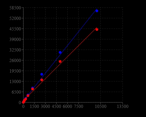
Cell number correlates with the luminescent signal. Different Jurkat cell number (2-fold dilution) was measured using the ReadiUse™ Rapid Luminometric ATP Assay Kit in a 96-well white plate using a ClarioStar plate reader (BMG Labtech). The kit can detect as low as 50 cells. There is a linear relationship (r2 >0.99) between the luminescent signal and cell number after 15 minutes or 2 hours incubation.  The half-life of luminescent signal is more than 2 hours.
Cell number correlates with the luminescent signal. Different Jurkat cell number (2-fold dilution) was measured using the ReadiUse™ Rapid Luminometric ATP Assay Kit in a 96-well white plate using a ClarioStar plate reader (BMG Labtech). The kit can detect as low as 50 cells. There is a linear relationship (r2 >0.99) between the luminescent signal and cell number after 15 minutes or 2 hours incubation. The half-life of luminescent signal is more than 2 hours.
Ordering information
Price
Unit size
100 Tests
1000 Tests
10000 Tests
Catalog Number
Quantity
Add to cart
Additional ordering information
Telephone1-800-990-8053
Fax1-800-609-2943
Emailsales@aatbio.com
InternationalSee distributors
Bulk requestInquire
Custom sizeInquire
ShippingStandard overnight for United States, inquire for international
Request quotation
Storage, safety and handling
H-phraseH303, H313, H333
Hazard symbolXN
Intended useResearch Use Only (RUO)
R-phraseR20, R21, R22
StorageFreeze (< -15 °C); Minimize light exposure
UNSPSC12352200
Platform

Luminescence microplate reader

Recommended plateSolid white
Example protocol

AT A GLANCE

Protocol Summary

  1. Prepare cells (samples) with test compounds (100 µL/96-well plate or 25 µL/384-well plate)
  2. Add equal volume of ready-to-use ReadiUse™ Rapid Luminometric ATP Assay Reagent (100 µL/96-well plate or 25 µL/384-well plate)
  3. Incubate at room temperature for 10 - 20 minutes
  4. Monitor the luminescence intensity

Important notes
To achieve the best results, it’s strongly recommended to use the white plates. Thaw all the kits components at room temperature before starting the experiment.

PREPARATION OF STANDARD SOLUTION

ATP standard

For convenience, use the Serial Dilution Planner: https://www.aatbio.com/tools/serial-dilution/21601

Optional, ATP is not provided.

Make 1 mM ATP stock solution with ddH2O or appropriate buffer, then perform serial dilution to achieve ATP concentrations ranging from 10 pM to 10 µM.

For guidelines on cell sample preparation, please visit
https://www.aatbio.com/resources/guides/cell-sample-preparation.html

SAMPLE EXPERIMENTAL PROTOCOL

Table 1. Layout of ATP Standards and test samples in a white 96-well microplate. SD=ATP Standard, BL=Blank Control, TS=Test Sample

BLBLTS TS
SD1SD1......
SD2SD2......
SD3SD3  
SD4SD4  
SD5SD5  
SD6SD6  
SD7 SD7  

Table 2. Reagent composition for each well

WellVolumeReagent
SD1-SD7100 µLSerial Dilution of ATP  (e.g. 10 pM to 10 µM)
BL100 µLATP Assay Buffer (Component A)
TS100 µLSample
  1. Treat cells (or samples) with test compounds by adding 10 µL of 10X compounds for a 96-well plate or 5 µL of 5X compounds for a 384-well plate in desired compound buffer. For blank wells (medium without the cells), add the corresponding amount of compound buffer.
  1. Incubate the cell plate in a 37 oC, 5% CO2 incubator for the desired period of time, such as 24, 48 or 96 hours.
  1. Add 100 µL/well (96-well plate) or 25 µL/well (384-well plate) of ReadiUse™ Rapid Luminometric ATP Assay Reagent.

  2. Incubate at room temperature for 10 - 20 minutes. Note: Aliquot and store the unused ReadiUse™ Rapid Luminometric ATP Assay Reagent at -20 oC, and avoid repeated freeze/thaw cycles.
  1. Monitor the luminescence intensity with a standard luminometer.
Citations
View all 15 citations: Citation Explorer
Palmitic Acid Exerts Anti-Tumorigenic Activities by Modulating Cellular Stress and Lipid Droplet Formation in Endometrial Cancer
Authors: Zhao, Ziyi and Wang, Jiandong and Kong, Weimin and Newton, Meredith A and Burkett, Wesley C and Sun, Wenchuan and Buckingham, Lindsey and O’Donnell, Jillian and Suo, Hongyan and Deng, Boer and others,
Journal: Biomolecules (2024): 601
Combining Cisplatin with Pinellia pedatisecta Schott Lipid-Soluble Extract Induces Tumor Immunogenic Cell Death in Cervical Cancer
Authors: Wang, Congwen and Zhang, Mingxing and Peng, Jing and Zhang, Meng and Lu, Chong and Qi, Xingling and Luo, Qingyan and Wang, Yumeng and Li, Guiling
Journal: Phytomedicine (2024): 155504
Homogeneous luminescent quantitation of cellular guanosine and adenosine triphosphates (GTP and ATP) using QT-LucGTP\&ATP assay
Authors: Kopra, Kari and Mahran, Randa and Yli-Hollo, Titta and Tabata, Sho and Vuorinen, Emmiliisa and Fujii, Yuki and Vuorinen, Iida and Ogawa-Iio, Aki and Hirayama, Akiyoshi and Soga, Tomoyoshi and others,
Journal: Analytical and Bioanalytical Chemistry (2023): 1--12
Guidelines for cell viability assays
Authors: Kamiloglu, Senem and Sari, Gulce and Ozdal, Tugba and Capanoglu, Esra
Journal: Food Frontiers (2020): 332--349
The Different Effects of Atorvastatin and Pravastatin on Cell Death and PARP Activity in Pancreatic NIT-1 Cells
Authors: Chen, Ya-Hui and Chen, Yi-Chun and Liu, Chin-San and Hsieh, Ming-Chia
Journal: Journal of Diabetes Research (2016)