mFluor™ Violet 540-Wheat Germ Agglutinin (WGA) Conjugate
| Price | |
| Catalog Number | |
| Unit Size | |
| Quantity |
| Telephone | 1-800-990-8053 |
| Fax | 1-800-609-2943 |
| sales@aatbio.com | |
| International | See distributors |
| Bulk request | Inquire |
| Custom size | Inquire |
| Shipping | Standard overnight for United States, inquire for international |
| Solvent | Water |
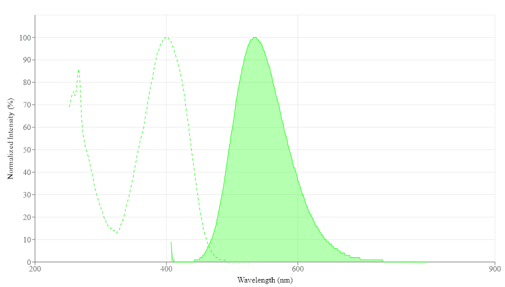
| Absorbance (nm) | 401 |
| Correction Factor (260 nm) | 1.326 |
| Correction Factor (280 nm) | 0.543 |
| Extinction coefficient (cm -1 M -1) | 180001 |
| Excitation (nm) | 402 |
| Emission (nm) | 535 |
| Quantum yield | 0.211 |
| H-phrase | H303, H313, H333 |
| Hazard symbol | XN |
| Intended use | Research Use Only (RUO) |
| R-phrase | R20, R21, R22 |
| Storage | Freeze (< -15 °C); Minimize light exposure |
| UNSPSC | 12171501 |
| Overview |
Absorbance (nm) 401 | Correction Factor (260 nm) 1.326 | Correction Factor (280 nm) 0.543 | Extinction coefficient (cm -1 M -1) 180001 | Excitation (nm) 402 | Emission (nm) 535 | Quantum yield 0.211 |
Platform
Fluorescence microscope
| Excitation | 402 nm |
| Emission | 535 nm |
| Recommended plate | Black wall/clear bottom |
Example protocol
PREPARATION OF STOCK SOLUTIONS
Unless otherwise noted, all unused stock solutions should be divided into single-use aliquots and stored at -20 °C after preparation. Avoid repeated freeze-thaw cycles
Add 500 µL of ddH2O into the powder form to make a 2 mg/mL stock solution.
Note: The reconstituted conjugate solution can be stored at 2-8 °C for short-term storage or at -20 °C for long-term storage.
PREPARATION OF WORKING SOLUTION
Add 5 µL of 200X WGA conjugate solution to 1 mL HHBS Buffer.
Note: The optimized staining concentration may be different with different cell lines. The recommended starting concentration is 5-10 µg/mL for live cells.
SAMPLE EXPERIMENTAL PROTOCOL
Warm the vial to room temperature centrifuge briefly before opening. Staining protocols vary with applications. Appropriate dilution of conjugates should be determined experimentally.
- Wash cells twice with a HHBS buffer.
Add 100 µL mFluor™ Violet 540-WGA working solution.
- Incubate cells with WGA working solution for 10-30 minutes at 37 °C.
- Wash cells twice with HHBS buffer.
Image cells on a fluorescence microscope using Ex/Em = 402/535 nm.
WGA conjugates can be also used to stain fixed cells.
Fix cells with 4% Formaldehyde in PBS.
Note: For fixed cell membrane staining, it is recommended to stain without the permeabilization step. A permeabilization step after fixation can facilitate staining intracellular compartments such as Golgi and Endoplasmic Reticulum (ER) structures.Add 100 µL mFluor™ Violet 540-WGA working solution.
- Incubate cells with WGA working solution for 10-30 minutes at room temperature.
- Wash cells twice with HHBS buffer.
Image cells on a fluorescence microscope using Ex/Em = 402/535 nm.
Spectrum
Spectral properties
| Absorbance (nm) | 401 |
| Correction Factor (260 nm) | 1.326 |
| Correction Factor (280 nm) | 0.543 |
| Extinction coefficient (cm -1 M -1) | 180001 |
| Excitation (nm) | 402 |
| Emission (nm) | 535 |
| Quantum yield | 0.211 |
Product Family
| Name | Excitation (nm) | Emission (nm) | Extinction coefficient (cm -1 M -1) | Quantum yield | Correction Factor (260 nm) | Correction Factor (280 nm) |
| mFluor™ Violet 450-Wheat Germ Agglutinin (WGA) Conjugate | 406 | 445 | 350001 | 0.811 | 0.338 | 0.078 |
| mFluor™ Violet 500-Wheat Germ Agglutinin (WGA) Conjugate | 410 | 501 | 250001 | 0.811 | 0.769 | 0.365 |
References
Authors: Tanida, Isei and Yamaguchi, Junji and Suzuki, Chigure and Kakuta, Soichiro and Uchiyama, Yasuo
Journal: Heliyon (2023): e17394
Authors: Klawonn, Isabell and Dunker, Susanne and Kagami, Maiko and Grossart, Hans-Peter and Van den Wyngaert, Silke
Journal: Microbial ecology (2023): 9-23
Authors: Montroni, Devis and Di Giosia, Matteo and Calvaresi, Matteo and Falini, Giuseppe
Journal: Molecules (Basel, Switzerland) (2022)
Authors: Buckle, Tessa and van der Wal, Steffen and van Willigen, Danny M and Aalderink, Germaine and KleinJan, Gijs H and van Leeuwen, Fijs W B
Journal: Theranostics (2020): 9890-9898
Authors: Yockey, Johnathan and Andres, Luke and Carson, Moleigh and Ory, Jeramia J and Reese, Amy J
Journal: mSphere (2019)
Authors: Hayashi, Keiko and Yoshida, Tomofumi and Hayano-Saito, Yuriko
Journal: Plant methods (2019): 159
Authors: de Wit, Sanne and Zeune, Leonie L and Hiltermann, T Jeroen N and Groen, Harry J M and Dalum, Guus van and Terstappen, Leon W M M
Journal: Cancers (2018)
Authors: Apfelthaler, C and Anzengruber, M and Gabor, F and Wirth, M
Journal: European journal of pharmaceutics and biopharmaceutics : official journal of Arbeitsgemeinschaft fur Pharmazeutische Verfahrenst (2017): 131-139
Authors: Lin, Shiqi and Yang, Ling and Chen, Gu and Li, Bing and Chen, Dingqiang and Li, Lin and Xu, Zhenbo
Journal: Microbial pathogenesis (2017): 285-291
Authors: de Fátima MenegociEugênio, Patrícia and Assunção, Nilson Antonio and Sciandra, Francesca and Aquino, Adriano and Brancaccio, Andrea and Carrilho, Emanuel
Journal: Electrophoresis (2016): 321-34
Application notes
Fluorescent Oligonucleotide Labeling Reagents
Monitoring of Mitochondrial Membrane Potential Changes in Live Cells Using JC-10
Selective Analysis of RNA in Live and Fixed Cells with StrandBrite RNA Green
Cell Loading Protocol For Fluorescent pH Indicator, BCECF-AM
FAQ
What dye works best for staining and tracking lysosomes in live cells for several hours?
How can I lyse my cells without lysing the nuclear membrane?
Do you have any dual-fluorescence nucleic acid stains that interact with both DNA and RNA?
Do you have any fixable mitochondria staining assay kits?