
Products
Services
Resources
Selection Guides
About
Lysosomes
Lysosomal dysfunction underlies a broad spectrum of pathologies—from inherited lysosomal storage diseases to neurodegeneration, cancer, and infectious disease. Researchers studying lysosomal biogenesis, autophagy flux, drug resistance mechanisms, or therapeutic targeting need reliable tools that report on lysosomal localization, pH dynamics, and membrane integrity without disrupting the processes under investigation.
AAT Bioquest's lysosome and autophagy probes address these needs across multiple experimental platforms. For example, our LysoBrite™ dyes, which exploit the lysosomal pH gradient for selective accumulation and pH-sensitive fluorescence enhancement, are available in spectral variants from blue to NIR for multiplexing flexibility. Additionally, our CytoFix™ Red reagent enables lysosomal tracking through fixation and downstream immunostaining workflows. Finally, our Cell Meter™ autophagy kits allow monitoring of autophagic activity in response to cellular stress, infection, or pharmacological intervention.
| Use for | Colors | |
|---|---|---|
| Live-Cell Staining | Labeling and tracking acidic lysosomes in live cells | Blue to near-infrared |
| Fixable Staining | Lysosome labeling that stays in place through fixation and immunostaining | Orange-red (Cy3/TRITC filter set) |
| Autophagy Assays | Detecting autophagosomes and mitophagy in live cells | Blue, green, and red |
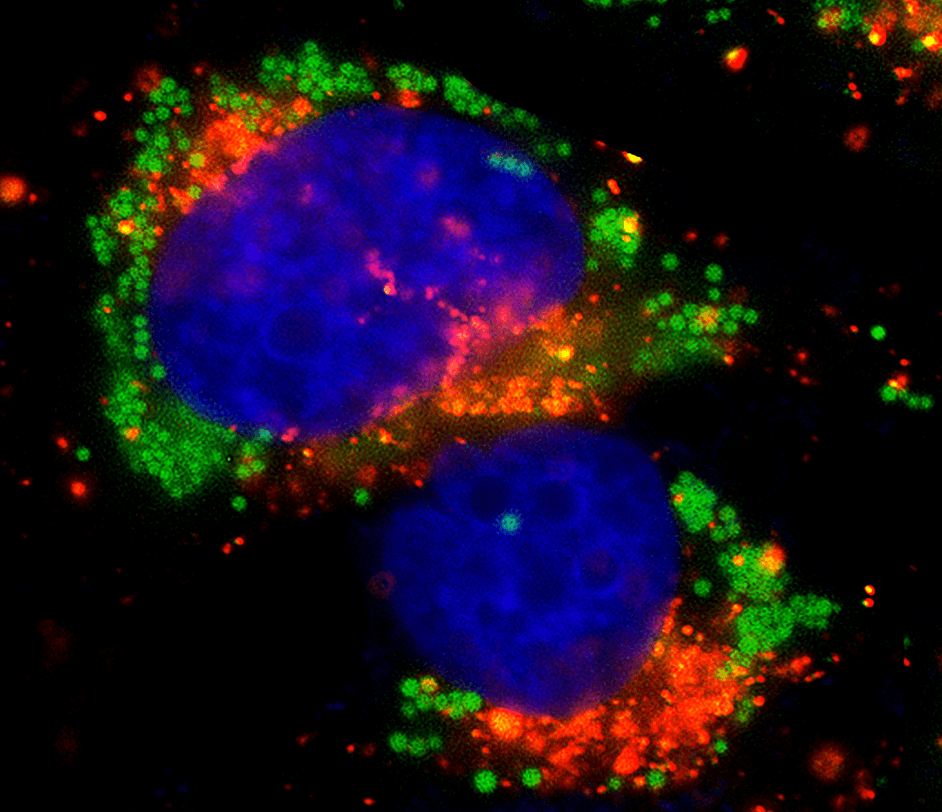 HeLa cells were incubated in 1X HBSS buffer with 5% serum to induce starvation. Following starvation, cells were treated with Autophagy Green™ (Catalog Number 23002) working solution for 20 minutes in a 37 °C, 5% CO2 incubator and then washed 3 times. Nuclei were labeled with Hoechst 33342 (Catalog Number 17530). Lysosomes were labeled with LysoBrite™ Orange (Catalog Number 22657).
LysoBrite™ dyes and Protonex™ Lyso probes are cell-permeable and accumulate in lysosomes and other acidic organelles of live cells.
LysoBrite™ dyes are cell-permeable probes that selectively accumulate in lysosomes and acidic organelles via the lysosomal pH gradient. At nanomolar concentrations, pH-sensitive fluorescence enhancement upon lysosomal entry generates high-contrast signal suitable for fluorescence imaging, high-content screening, microplate fluorometry, or flow cytometry.
LysoBrite™ dyes span blue to near-infrared wavelengths for multiplexing with other fluorescent probes. LysoBrite™ Orange, Red, Deep Red, and NIR variants provide stable lysosomal labeling suitable for imaging-based studies where lysosomal behavior is examined in the context of processes such as cell adhesion, chemotaxis, multidrug resistance, cell viability, apoptosis, and cytotoxicity. LysoBrite™ Red DND-99 (Catalog Number 22647) retains signal after aldehyde fixation for co-localization studies. LysoBrite™ dyes are the core components of Cell Navigator® Lysosome Staining Kits, which add an optimized cell-labeling protocol and provide materials for 500 tests.
pH-sensitive fluorescence for specific staining with minimal background | Photostable signal for extended imaging |
Minimal cytotoxicity with a no-wash imaging protocol | Works in proliferating and non-proliferating cells, suspension and adherent formats |
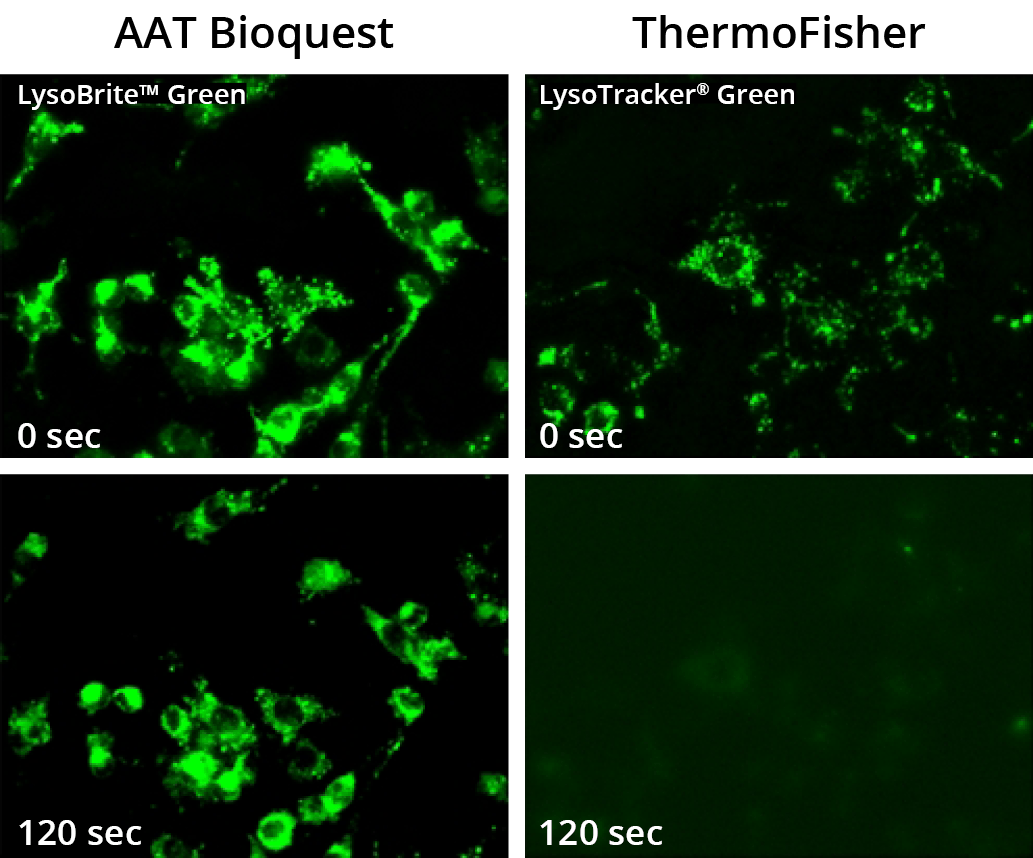 Images of HeLa cells stained with AAT Bioquest's LysoBrite™ Green (Catalog Number 22643) and ThermoFisher's LysoTracker® Green DND-26 in a Costar black wall/clear bottom 96-well microplate. Samples were continuously illuminated for 120 seconds and the signals were compared before and after exposure using a Keyence fluorescence microscope.
Protonex™ Lyso-Red 670 (Catalog Number 21241) and Protonex™ Lyso-NIR 770 (Catalog Number 21242) are cell-permeable pH probes that localize in acidic organelles such as lysosomes. Lyso-Red 670 is weakly fluorescent at alkaline pH and brightens as acidity increases, for real-time tracking of lysosomal dynamics. Lyso-NIR 770 is read with a common Cy7 filter set, and its red-shifted excitation and emission give lower background than conventional pH sensors.
CytoFix™ Red maintains lysosomal localization through fixation—critical for experiments requiring immunostaining, long-term storage, or batch processing. Enhanced cellular retention preserves fluorescence signal for at least one week post-fixation, enabling longitudinal lysosomal tracking studies.
CytoFix™ Red multiplexes with GFP, other fluorescent conjugates, and organelle stains. It is compatible with suspension and adherent cells across fluorescence microscopy, flow cytometry, and high-content platforms.
High lysosomal staining efficiency | Signal retained for at least one week after fixation |
Minimal hands-on time | Multiplexes with GFP, other fluorescent conjugates and organelle stains |
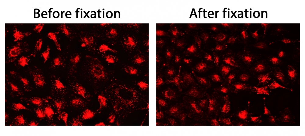 The fluorescence images of HeLa cells stained with CytoFix™ LysoRed. Image was acquired before (Left) and after (Right) fixation with 4% formaldehyde solution for 20 minutes at RT. The cells were imaged using fluorescence microscope with a Cy3/TRITC filter.
Autophagy is a lysosomal degradation pathway that sequesters cytoplasmic cargo into double-membrane autophagosomes, which subsequently fuse with lysosomes for content digestion and recycling. As a protective mechanism against cellular stress, infection, and metabolic dysfunction, autophagy is a key target in cancer, neurodegeneration, and infectious disease research.
Cell Meter™ autophagy kits employ Autophagy Blue™, Autophagy Green™, or Autophagy Super Blue™ as specific autophagosome markers for the direct detection of autophagy in detached and attached live cells. The Cell Meter™ Mitochondrial Autophagy Imaging Kit uses Mitophagy Red™, which stains mitochondria and translocates to lysosomes upon induction of mitophagy.
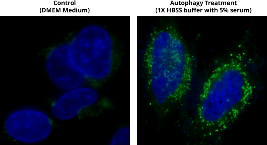 Autophagy Green™ labeled vesicles are induced by starvation in HeLa cells. HeLa cells were incubated in a regular DMEM medium (Control) or in 1X HBSS buffer with 5% serum (Autophagy Treatment) for 16 hours. Both control cells and starved cells were incubated with Autophagy Green™ working solution for 20 minutes in a 37°C, 5% CO2 incubator, and then washed 3 times with wash buffer. Cells were imaged immediately under a fluorescence microscope with a FITC channel (green). Cell nuclei were stained with Hoechst 33342 (Catalog Number 17530).
This document (01.0085.211015r2) was last updated on Wed Nov 11 2026. All trademarks and registered trademarks mentioned herein are the property of their respective owners.