
Products
Services
Resources
Selection Guides
About
iFluor® 488 succinimidyl ester
iFluor® 488 succinimidyl ester is a green-fluorescent, 488 nm excitable dye that is suitable for fluorescence imaging and flow cytometry applications.
- Bioconjugation Ready: Label the primary amines (R-NH2) of proteins, antibodies, amine-modified oligonucleotides and other amine-containing biomolecules
- Improved Photostability and Brightness: Compared to FITC, iFluor® 488 conjugates exhibit higher fluorescence intensity and are less susceptible to photobleaching
- Platform Validated: Ideally suited for fluorescence microscopy, flow cytometry and spectral flow cytometry
- Spectral Compatibility: iFluor® 488 has spectral properties similar to Alexa Fluor® 488 and FITC, allowing it to easily slot into existing workflows
 using fluorescence microscope with a FITC filter set (Green). The cells were fixed in 4% formaldehyde, co-labeled with mitochondria dye [MitoLite™ Red FX600](/products/mitolite-red-fx600) (Red) and [Nuclear Blue™ DCS1](/products/nuclear-blue-dcs1-5-mm-dmso-solution) (Blue).](/_next/image?url=https%3A%2F%2Fimages.aatbio.com%2Fproducts%2Ffigures-and-data%2Fifluor-488-succinimidyl-ester%2Ffigure-for-ifluor-488-succinimidyl-ester_Pk16S.jpeg&w=1920&q=100)

| Catalog | Size | Price | Quantity |
|---|---|---|---|
| 1023 | 1 mg | Price | |
| 71023 | 100 ug | Price | |
| 71503 | 5 mg | Price | |
| 71553 | 10 mg | Price |
Overview
Bioconjugation Ready
iFluor® 488 succinimidyl ester (NHS ester) is an amine-reactive fluorescent dye that can be used to label the primary amines on proteins and antibodies (such as lysine residues), as well as other amine-containing biomolecules. For instance, it can be conjugated to a secondary antibody as part of a signal amplification system (Figure 1), or it can be conjugated to an amine-modified oligonucleotide as part of a fluorescence in situ hybridization (FISH) experiment (Figure 2).
 using fluorescence microscope with a FITC filter set (Green). The cells were fixed in 4% formaldehyde, co-labeled with mitochondria dye [MitoLite™ Red FX600](/products/mitolite-red-fx600) (Red) and [Nuclear Blue™ DCS1](/products/nuclear-blue-dcs1-5-mm-dmso-solution) (Blue).](https://images.aatbio.com/products/figures-and-data/ifluor-488-succinimidyl-ester/figure-for-ifluor-488-succinimidyl-ester_Pk16S.jpeg)
) for spectral reference.
(Bottom) Flow cytometry analysis of PBMCs stained with [iFluor® 488 Anti-human CD24](/products/ifluor-488-anti-human-cd24-antibody-hi45) (Clone: HI45) conjugate. The fluorescence signal was monitored using a Cytek Aurora spectral flow cytometer in the B2-A channel.](https://images.aatbio.com/products/figures-and-data/ifluor-488-succinimidyl-ester/figure-for-ifluor-488-succinimidyl-ester_gaCHc.png)
Figure 2. (Top) Spectral signature of iFluor® 488 dye. Data were acquired on a 4-laser Cytek Aurora using normal human peripheral blood cells stained with Anti-human CD4 (Clone: SK3) conjugated to iFluor® 488 dye (Cat. No. 10042050) for spectral reference. (Bottom) Flow cytometry analysis of PBMCs stained with iFluor® 488 Anti-human CD24 (Clone: HI45) conjugate. The fluorescence signal was monitored using a Cytek Aurora spectral flow cytometer in the B2-A channel.
Conjugation of the dye to a protein or an antibody of choice typically involves incubation at room temperature for 30 to 60 minutes. Afterwards, purification with a size exclusion column (such as Sephadex G-25 or P-6-DG) is recommended.
For a no purification variant of iFluor® 488 succinimidyl ester, please see Catalog 1255.
Additionally, it is recommended to characterize the resulting conjugates by calculating the degree of labeling (DOL), also known as the degree of substitution (DOS). This can be performed using a Degree of Labeling Calculator.
Photostability and Brightness
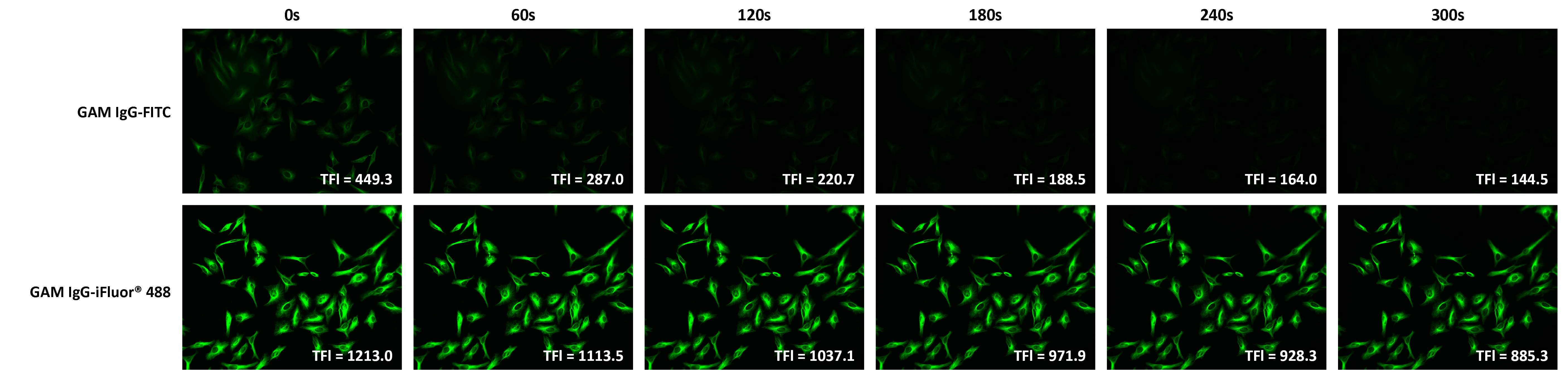
iFluor® 488 has excellent photostability and superior brightness when compared to classic green-fluorescent dyes, such as fluorescein (FITC). In a comparative experiment, iFluor® 488 and FITC were conjugated to goat anti-mouse IgG and used to bind mouse anti-tubulin. It was found that the fluorescence intensity loss of FITC was more than double that of iFluor® 488 (68% vs 27%) over the same period of time (Figure 3). Thus, iFluor® 488 presents significant advantages in experiments where photostability or long exposure times are of concern.
, and nuclei were stained with [Nuclear Red DCS1](/products/nuclear-red-dcs1-5-mm-dmso-solution).](https://images.aatbio.com/products/figures-and-data/ifluor-488-succinimidyl-ester/figure-for-ifluor-488-succinimidyl-ester_fubUW.jpg)
Additionally, the fluorescence of iFluor® 488 is not affected by pH (4-10). This pH insensitivity is a major improvement over fluorescein, which emits its maximum fluorescence only at pH above 9.
Platform Validated
iFluor® 488 has been validated across multiple instrument platforms, including fluorescence microscopy, flow cytometry and spectral flow cytometry.
Fluorescence Microscopy
In fluorescence microscopy, iFluor® 488 is efficiently excited by the 488 nm laser line and is fully compatible with standard FITC filter sets, providing strong green emission with minimal background (Figure 4).
 Figure 4. Images of HeLa cells captured using Keyence X710 fluorescence microscope. TFI represents total fluorescence intensity as calculated by ImageJ software.
In fluorescence microscopy, iFluor® 488 is efficiently excited by the 488 nm laser line and is fully compatible with standard FITC filter sets, providing strong green emission with minimal background (Figure 4).
Flow Cytometry
In flow cytometry, iFluor® 488 can be reliably detected in the FITC channel (Figure 5), offering a convenient replacement for traditional green fluorophores like FITC and Alexa Fluor® 488.
 Figure 5. Fluorescence In Situ Hybridization of Fluorescein and iFluor® 488 labelled Telomere probes in metaphase HeLa cells.
In flow cytometry, iFluor® 488 can be reliably detected in the FITC channel (Figure 5), offering a convenient replacement for traditional green fluorophores like FITC and Alexa Fluor® 488.

Spectral Flow Cytometry
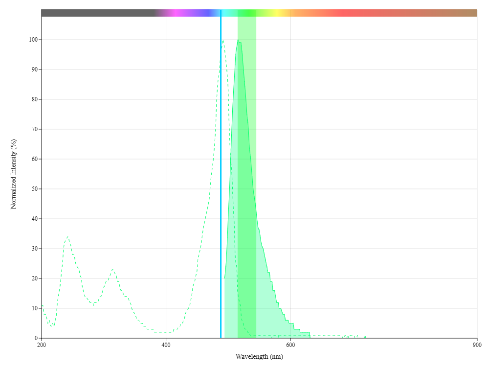
iFluor® 488 is compatible with next generation spectral flow cytometers, such as the Cytek Aurora, allowing for expanded multiplexing capabilities. PBMCs stained with iFluor® 488 anti-human CD24 HI45 conjugate showed strong signal in the Aurora’s B2-A channel (Figure 6, Bottom). Spectral analysis of CD4-labeled human peripheral blood cells further confirmed the unique emission profile of iFluor® 488 on a 4-laser Aurora system (Figure 6, Top).
An example panel designed with iFluor® dye technology can be found here. To design a custom panel for your specific instrument, please see the Spectral Flow Cytometry Panel Builder Tool.
 Figure 6. iFluor® 488 spectrum with 488 nm excitation laser and FITC filter.
iFluor® 488 is compatible with next generation spectral flow cytometers, such as the Cytek Aurora, allowing for expanded multiplexing capabilities. PBMCs stained with iFluor® 488 anti-human CD24 HI45 conjugate showed strong signal in the Aurora’s B2-A channel (Figure 6, Bottom). Spectral analysis of CD4-labeled human peripheral blood cells further confirmed the unique emission profile of iFluor® 488 on a 4-laser Aurora system (Figure 6, Top).
An example panel designed with iFluor® dye technology can be found here. To design a custom panel for your specific instrument, please see the Spectral Flow Cytometry Panel Builder Tool.
Spectral Compatibility
iFluor® 488 (ex / em = 491 / 516 nm) has spectral properties similar to Alexa Fluor® 488 (ex / em = 494 / 517 nm), allowing for straightforward implementation into existing workflows (Figure 7, Figure 8).
 (Green, Left) or Alexa Fluor® 488 goat anti-mouse IgG conjugate (Green, Right), respectively. Cell nuclei were stained with [Hoechst 33342](/products/hoechst-33342-ultrapure-grade) (Blue).](https://images.aatbio.com/products/figures-and-data/ifluor-488-succinimidyl-ester/figure-for-ifluor-488-succinimidyl-ester_Z7U1g.jpg)
 of iFluor® 488 and Alexa Fluor® 488 spectra](https://images.aatbio.com/products/figures-and-data/ifluor-488-succinimidyl-ester/figure-for-ifluor-488-succinimidyl-ester_rPG5i.png)
Custom Conjugation Service
We provide custom conjugation services for all of our iFluor® dyes. Please see here for more information.
Useful Resources
Alexa Fluor® is a trademark or registered trademark of ThermoFisher Scientific.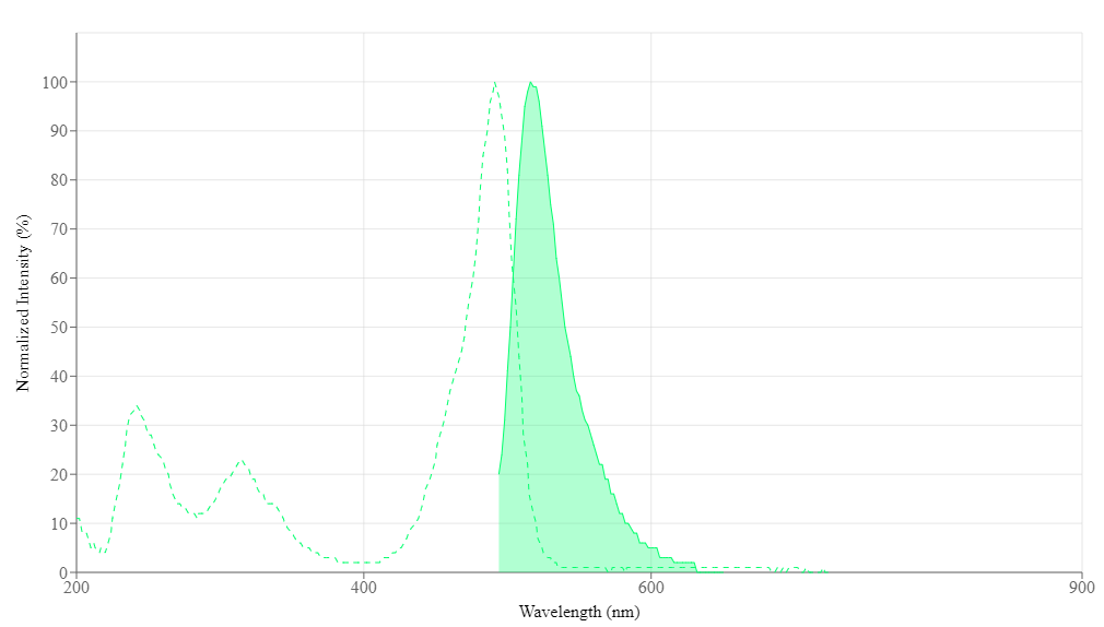
Physical properties
| Molecular weight | 945.07 |
| Solvent | DMSO |
Spectral properties
| Correction factor (260 nm) | 0.21 |
| Correction factor (280 nm) | 0.11 |
| Extinction coefficient (cm -1 M -1) | 75000 1 |
| Excitation (nm) | 491 |
| Emission (nm) | 516 |
| Quantum yield | 0.9 1 |
Usage and storage
| Intended use | Research Use Only (RUO) |
| Storage | Freeze (< -15 °C); Minimize light exposure |
Contact us
| Telephone | |
| Fax | |
| sales@aatbio.com | |
| International | See distributors |
| Bulk request | Inquire |
| Custom size | Inquire |
| Technical support | Contact us |
| Request quotation | Request |
| Purchase order | Send to sales@aatbio.com |
| Shipping | Standard overnight for United States, inquire for international |
Page updated on October 19, 2026