Cell Meter™ Fluorimetric Intracellular Total ROS Activity Assay Kit*Green Fluorescence*
Overview | ![]() ![]() |
Platform
Fluorescence microscope
Excitation | FITC filter |
Emission | FITC filter |
Recommended plate | Black wall/clear bottom |
Fluorescence microplate reader
Excitation | 490 nm |
Emission | 525 nm |
Cutoff | 515 nm |
Recommended plate | Black wall/clear bottom |
Instrument specification(s) | Bottom read mode |
Components
Component A: Amplite™ ROS Green | 1 vial |
Component B: Assay Buffer | 1 bottle (20 mL) |
Component C: DMSO | 1 vial (200 µL) |
Example protocol
AT A GLANCE
Protocol summary
- Prepare cells in growth medium
- Add Amplite™ ROS Green working solution (100 µL/well for a 96- well plate or 25 µL/well for a 384-well plate)
- Stain the cells at 37°C for 60 minutes
- Treat the cells with test compounds to induce ROS
- Monitor the fluorescence increase (bottom read mode) at Ex/Em= 490/525 nm (Cutoff = 515 nm) or fluorescence microscope with FITC filter set
Important notes
Thaw all the kit components at room temperature before starting the experiment.
PREPARATION OF STOCK SOLUTION
1. Amplite™ ROS Green stock solution (500X):
Add 40 µL of DMSO (Component C) into the vial of Amplite™ ROS Green (Component A) and mix well to make 500X Amplite™ ROS Green stock solution. Protect from light. Note: 20 µL of 500X Amplite™ ROS Green stock solution is enough for 1 plate. For storage, seal tubes tightly.
PREPARATION OF WORKING SOLUTION
Add 20 µL of 500X Amplite™ ROS Green stock solution into 10 mL of Assay Buffer (Component B) and mix well to make Amplite™ ROS Green working solution. Note: This Amplite™ ROS Green working solution is stable for at least 2 hours at room temperature.
For guidelines on cell sample preparation, please visit
https://www.aatbio.com/resources/guides/cell-sample-preparation.html
SAMPLE EXPERIMENTAL PROTOCOL
- Add 100 µL/well (96-well plate) or 25 µL/well (384-well plate) of Amplite™ ROS Green working solution into the cell plate.
- Incubate the cells in a 5% CO2, 37°C incubator for one hour.
- Treat cells with 20 µL of 11X test compounds (96-well plate) or 10 µL of 6X test compounds (384-well plate) in your desired buffer (such as PBS or HHBS). For control wells (untreated cells), add the corresponding amount of compound buffer.
- To induce ROS, incubate the cell plate at room temperature or in a 5% CO2, 37°C incubator for at least 15 minutes or a desired period of time (30 minutes for Hela cells treated with 1 mM H2O2).
- Monitor the fluorescence increase with a fluorescence microplate reader (bottom read mode) at Ex/Em = 490/525 nm (Cutoff = 515 nm) or observe cells using a fluorescence microscope with FITC filter set.
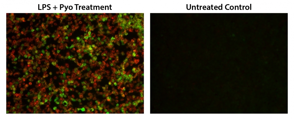
Images
Citations
Authors: Jiang, Xuan and Zhang, Xin and Fu, Chao and Zhao, Ruili and Jin, Tianming and Liu, Mengyue and Pan, Chenhao and Li, Liu An and Ma, Jifei and Yu, Enyuan and others,
Journal: The Protein Journal (2021): 1--12
Authors: Tang, Jingshu and Kang, Yuying and Zhou, Yujun and Li, Xinnan and Lan, Jiaqi and Wu, Lei and Feng, Xinhong and Peng, Ying
Journal: Neurobiology of Disease (2021): 105315
Authors: Shimoda, Ayumu and Tanabe, Takemi and Sato, Tsubasa and Nedachi, Taku
Journal: Bioscience, Biotechnology, and Biochemistry (2021): 2103--2112
Authors: Teng, Yu-Ning and Wang, Charles CN and Liao, Wei-Chieh and Lan, Yu-Hsuan and Hung, Chin-Chuan
Journal: Molecules (2020): 247
Authors: Lakshmi, Sowmya P and Reddy, Aravind T and Kodidhela, Lakshmi Devi and Varadacharyulu, N Ch
Journal: Life Sciences (2020): 118260
Authors: Bayne, Mitchell and Alvarsson, Alexandra and Devarakonda, Kavya and Li, Rosemary and Jimenez-Gonzalez, Maria and Garibay, Darline and Conner, Kaetlyn and Varghese, Merina and Serasinghe, Madhavika N and Chipuk, Jerry E and others,
Journal: JCI insight (2020)
Authors: Liu, Feng and Wang, Wencheng and Xia, Yunqiu and Chen, Xuehong and Han, Yantao and Miao, Desen and Zhang, Deyan and Lv, Hong and Yang, Desheng and Zhang, Daisong and others,
Journal: Journal of King Saud University-Science (2020)
Authors: Takeda, Tomoya and Doiyama, Sota and Azumi, Junya and Shimada, Yasuhiro and Tokuji, Yoshihiko and Yamaguchi, Hiroaki and Nagata, Kosuke and Sakamoto, Naoya and Aso, Hisashi and Nakamura, Takashi
Journal: Scientific reports (2019): 1--17
Authors: Kulkarni, Nishant S and Parvathaneni, Vineela and Shukla, Snehal K and Barasa, Leonard and Perron, Jeanette C and Yoganathan, Sabesan and Muth, Aaron and Gupta, Vivek
Journal: European Journal of Pharmaceutical Sciences (2019)
Authors: Hayashi, Nobuya and Miyamaru, Yukie and Aijima, Reona and Yamashita, Yoshio
Journal: IEEE Transactions on Plasma Science (2018): 1--7
References
Authors: Magalhaes LM, Lucio M, Segundo MA, Reis S, Lima JL.
Journal: Talanta (2009): 1219
Authors: Amaral S, Oliveira PJ, Ramalho-Santos J.
Journal: Curr Diabetes Rev (2008): 46
Authors: Murray BK, Ohmine S, Tomer DP, Jensen KJ, Johnson FB, Kirsi JJ, Robison RA, O'Neill KL.
Journal: J Virol Methods (2008): 74
Authors: Starkov AA., undefined
Journal: Ann N Y Acad Sci (2008): 37
Authors: Wada M., undefined
Journal: Yakugaku Zasshi (2008): 1031
Authors: Perrone GG, Tan SX, Dawes IW.
Journal: Biochim Biophys Acta (2008): 1354
Authors: Armstrong JS, Whiteman M.
Journal: Methods Cell Biol (2007): 355
Authors: Zhang W, Wang M, Xie HY, Zhou L, Meng XQ, Shi J, Zheng S.
Journal: Transplant Proc (2007): 1332
Authors: Gorlach A, Kietzmann T.
Journal: Methods Enzymol (2007): 421
Authors: Afonso V, Champy R, Mitrovic D, Collin P, Lomri A.
Journal: Joint Bone Spine (2007): 324
Application notes
Fluorimetric detection of ATPase and protein phosphatase activities by monitoring formation of inorganic phosphate
β-adrenoceptors are upregulated in human melanoma and their activation releases pro-tumorigenic cytokines and metalloproteases in melanoma cell lines
EphA2 Induces Metastatic Growth Regulating Amoeboid Motility and Clonogenic Potential in Prostate Carcinoma Cells
Matrix Remodeling Maintains Embryonic Stem Cell Self-Renewal by Activating Stat3
Selective Detection of Pyrophosphate Using a Fluorogenic Pyrophosphate Sensor