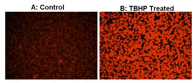
Cell Meter™ Fluorimetric Intracellular Total ROS Activity Assay Kit*Orange Fluorescence*
Overview | ![]() ![]() |
Platform
Flow cytometer
Excitation | 488 nm or 532 nm laser |
Emission | 575/26 nm filter |
Instrument specification(s) | PE channel |
Fluorescence microscope
Excitation | TRITC filter |
Emission | TRITC filter |
Recommended plate | Black wall/clear bottom |
Fluorescence microplate reader
Excitation | 540 nm |
Emission | 570 nm |
Cutoff | 550 nm |
Recommended plate | Black wall/clear bottom |
Instrument specification(s) | Bottom read mode |
Components
Component A: ROS Brite™ 570 | 1 vial |
Component B: Assay Buffer | 1 bottle (20 mL) |
Component C: DMSO | 1 vial (100 µL) |
Example protocol
AT A GLANCE
Protocol A summary (Fluorescence microplate reader, fluorescence microscope)
- Prepare cells in growth medium
- Treat the cells with test compounds to induce ROS
- Add ROS Brite™ 570 working solution (100 µL/well for a 96-well plate or 25 µL/well for a 384-well plate)
- Stain the cells at 37 °C for 30 - 60 minutes
- Monitor the fluorescence increase (bottom read mode) at Ex/Em= 540/570 nm (Cutoff = 550 nm) or fluorescence microscope with TRITC filter set
Protocol B summary (Flow cytometer)
- Prepare cells in growth medium
- Treat cells with test compounds to induce ROS
- Incubate ROS Brite™ 570 with the cells for 30 - 60 minutes
- Monitor the fluorescence intensities using flow cytometer with FL2 channel
Important notes
Thaw all the kit components at room temperature before starting the experiment.
PREPARATION OF STOCK SOLUTION
1. ROS Brite™ 570 stock solution (500X):
Add 40 µL of DMSO (Component C) into the vial of ROS Brite™ 570 (Component A) and mix well to make 500X ROS Brite™ 570 stock solution. Protect from light. Note: 20 µL of 500X ROS Brite™ 570 stock solution is enough for 1 plate. For flow cytometer, 500X ROS Brite™ 570 stock solution can be diluted by 5 folders to 100X in DMSO for convenience. For storage, seal tubes tightly.
PREPARATION OF WORKING SOLUTION
Add 20 µL of 500X ROS Brite™ 570 stock solution into 10 mL of Assay Buffer (Component B) and mix well to make ROS Brite™ 570 working solution. Note: This ROS Brite™ 570 working solution is stable for at least 2 hours at room temperature.
For guidelines on cell sample preparation, please visit
https://www.aatbio.com/resources/guides/cell-sample-preparation.html
SAMPLE EXPERIMENTAL PROTOCOL
For Protocol A:
- Treat cells with 10 µL of 10X test compounds (96-well plate) or 5 µL of 5X test compounds (384-well plate) in your desired buffer (such as PBS or HHBS). For control wells (untreated cells), add the corresponding amount of compound buffer.
- To induce ROS, incubate the cell plate at room temperature or in a 5% CO2, 37 °C incubator for a desired period of time (for example: 30 minutes treatment for Hela cells with 100 µM tert-butyl hydroperoxide (TBHP)).
- Add 100 µL/well (96-well plate) or 25 µL/well (384-well plate) of ROS Brite™ 570 working solution into the cell plate.
- Incubate the cells in a 5% CO2, 37 °C incubator for 30 min to 60 minutes.
- Monitor the fluorescence increase with a fluorescence microplate reader (bottom read mode) at Ex/Em = 540/570 nm (Cutoff = 550nm) or observe cells using a fluorescence microscope with TRITC filter set.
For Protocol B:
- Prepare cells at the density from 5 × 10⁵ to 1 × 10⁶ cells/mL. Note: Each cell line should be evaluated on the individual basis to determine the optimal cell density for apoptosis induction.
- Treat cells with test compounds in your desired buffer (such as PBS or HHBS). For control wells (untreated cells), add the corresponding amount of compound buffer.
- To induce ROS, incubate the cell plate at room temperature or in a 5% CO2, 37 °C incubator for at least 30 minutes or a desired period of time (30 minutes for Hela cells treated with 100 µM tert-butyl hydroperoxide (TBHP)).
- Add 1 µL/mL cells of 500X ROS Brite™ 570 stock solution or 5 µL/mL cells of 100X ROS Brite™ 570 stock solution to cells medium.
- Incubate the cells in a 5% CO2, 37 °C incubator for 30 to 60 minutes.
- Monitor the fluorescence intensity using a flow cytometer with FL2 channel.
Images
Citations
Authors: Fan, Chunlan and Qiao, Yuan and Tang, Minke
Journal: Drug design, development and therapy (2017): 3343
Authors: Fan, Chunlan and Qiao, Yuan and Tang, Minke
Journal: Drug Design, Development and Therapy (2017): 3343
Authors: Ma, Jianwei and Hiratsuka, Takahiro and Etoh, Tsuyoshi and Akada, Junko and Fujishima, Hajime and Shiraishi, Norio and Yamaoka, Yoshio and Inomata, Masafumi
Journal: Journal of Gastroenterology and Hepatology (2017)
Authors: Zhang, J and Shi, HS and Liu, JQ and Yu, T and Shen, ZH and Ye, JD
Journal: Journal of Materials Chemistry B (2015): 8782--8795
Authors: Patrick, Ping and Price, Tulin O and Diogo, Ana L and Sheibani, Nader and Banks, William A and Shah, Gul N
Journal: Journal of endocrinology and diabetes (2015)
Authors: Kawamura, Kazuyuki and Kuroda, Yukiko and Sogo, Masako and Fujimoto, Miki and Inui, Toshio and Mitsui, Takao
Journal: Biochemical and biophysical research communications (2014): 181--185
Authors: Lakshmi, Sowmya P and Reddy, Aravind T and Zhang, Yingze and Sciurba, Frank C and Mallampalli, Rama K and Duncan, Steven R and Reddy, Raju C
Journal: Journal of Biological Chemistry (2014): 6383--6393
Authors: Nomura, Johji and Busso, Nathalie and Ives, Annette and Matsui, Chieko and Tsujimoto, Syunsuke and Shirakura, Takashi and Tamura, Mizuho and Kobayashi, Tsunefumi and So, Alex and er , undefined and Yamanaka, Yoshihiro
Journal: Scientific reports (2014): 4554
Authors: Shah, Gul N and Morofuji, Yoichi and Banks, William A and Price, Tulin O
Journal: Biochemical and biophysical research communications (2013): 354--358
Authors: Berghea, Florian and Berghea, Camelia Elena and Abobului, Mihai
Journal: Internal Medicine : 11--15
References
Authors: Magalhaes LM, Lucio M, Segundo MA, Reis S, Lima JL.
Journal: Talanta (2009): 1219
Authors: Amaral S, Oliveira PJ, Ramalho-Santos J.
Journal: Curr Diabetes Rev (2008): 46
Authors: Murray BK, Ohmine S, Tomer DP, Jensen KJ, Johnson FB, Kirsi JJ, Robison RA, O'Neill KL.
Journal: J Virol Methods (2008): 74
Authors: Starkov AA., undefined
Journal: Ann N Y Acad Sci (2008): 37
Authors: Wada M., undefined
Journal: Yakugaku Zasshi (2008): 1031
Authors: Perrone GG, Tan SX, Dawes IW.
Journal: Biochim Biophys Acta (2008): 1354
Authors: Armstrong JS, Whiteman M.
Journal: Methods Cell Biol (2007): 355
Authors: Zhang W, Wang M, Xie HY, Zhou L, Meng XQ, Shi J, Zheng S.
Journal: Transplant Proc (2007): 1332
Authors: Gorlach A, Kietzmann T.
Journal: Methods Enzymol (2007): 421
Authors: Afonso V, Champy R, Mitrovic D, Collin P, Lomri A.
Journal: Joint Bone Spine (2007): 324
Application notes
Fluorimetric detection of ATPase and protein phosphatase activities by monitoring formation of inorganic phosphate
β-adrenoceptors are upregulated in human melanoma and their activation releases pro-tumorigenic cytokines and metalloproteases in melanoma cell lines
EphA2 Induces Metastatic Growth Regulating Amoeboid Motility and Clonogenic Potential in Prostate Carcinoma Cells
Matrix Remodeling Maintains Embryonic Stem Cell Self-Renewal by Activating Stat3
Selective Detection of Pyrophosphate Using a Fluorogenic Pyrophosphate Sensor