Cell Meter™ Intracellular Fluorimetric Hydrogen Peroxide Assay Kit *Green Fluorescence*
Price | |
Catalog Number | |
Unit Size | |
Quantity |
Telephone | 1-800-990-8053 |
Fax | 1-800-609-2943 |
sales@aatbio.com | |
International | See distributors |
Bulk request | Inquire |
Custom size | Inquire |
Shipping | Standard overnight for United States, inquire for international |
Excitation (nm) | 498 |
Emission (nm) | 517 |
H-phrase | H303, H313, H333 |
Hazard symbol | XN |
Intended use | Research Use Only (RUO) |
R-phrase | R20, R21, R22 |
UNSPSC | 12171501 |
Overview | ![]() ![]() |
Excitation (nm) 498 | Emission (nm) 517 |
Platform
Fluorescence microscope
Excitation | FITC channel |
Emission | FITC channel |
Recommended plate | Black wall/clear bottom |
Fluorescence microplate reader
Excitation | 490 nm |
Emission | 525 nm |
Cutoff | 515 nm |
Recommended plate | Black wall/clear bottom |
Instrument specification(s) | Bottom read mode |
Components
Example protocol
AT A GLANCE
Protocol summary for Solution Assay
- Prepare and add Amplite™ Green Peroxide Sensor working solution (50 µL)
- Add H2O2 standards or test samples (50 µL)
- Incubate at room temperature for 15 - 60 minutes
- Read fluorescence intensity at Ex/Em = 490/525 nm (Cutoff = 515 nm)
Important notes
Thaw all the kit components at room temperature before starting the experiment.
Protocol summary for Live Cell Assay
- Prepare cells in growth medium
- Stain cells with Amplite™ Green Peroxide Sensor working solution and incubate for your desired period of time
- Treat cells with test compounds
- Monitor fluorescence intensity at Ex/Em = 490/525 nm (Cutoff = 515 nm) with bottom read mode
Important notes
Amplite™ Green Peroxide Sensor can be loaded passively into living cells and report the micromolar changes in intracellular H2O2 concentrations. The following is a suggested microscope imaging protocol that can be modified to meet specific research needs.
PREPARATION OF STOCK SOLUTION
1. Amplite™ Green Peroxide Sensor stock solution (250X):
Add 50 µL of DMSO (Component D) into the vial of Amplite™ Green Peroxide Sensor (Component A) to make 250X Amplite™ Green Peroxide Sensor stock solution. Protect from light.
2. H2O2 standard solution (20 mM):
Add 22.7 µL of 3% H2O2 (0.88 M, Component B) into 977 µL of Assay Buffer (Component C) to make 20 mM H2O2 standard solution. Note: The diluted H2O2 standard solution is not stable. The unused portion should be discarded.
PREPARATION OF STANDARD SOLUTION
For convenience, use the Serial Dilution Planner: https://www.aatbio.com/tools/serial-dilution/11503
Add 50 µL of 20 mM H2O2 standard solution into 950 µL of Assay Buffer (Component C) to get 1000 µM H2O2 standard solution. Take 1000 µM H2O2 standard solution and perform 1:3 serial dilutions to get serially diluted H2O2 standards (HS1 - HS7) with Assay Buffer (Component C).
PREPARATION OF WORKING SOLUTION
Add 20 μL of 250X Amplite™ Green Peroxide Sensor stock solution into 5 mL of Assay Buffer (Component C) to make Amplite™ Green Peroxide Sensor working solution.
For guidelines on cell sample preparation, please visit
https://www.aatbio.com/resources/guides/cell-sample-preparation.html
SAMPLE EXPERIMENTAL PROTOCOL
Run H2O2 assay in supernatants reaction:
Table 1. Layout of H2O2 standards and test samples in a solid black 96-well microplate. HS= H2O2 Standards (HS1 - HS7, 300 to 0.3 µM); BL=Blank Control; TS=Test Samples
BL | BL | TS | TS |
HS1 | HS1 | ... | ... |
HS2 | HS2 | ... | ... |
HS3 | HS3 | ||
HS4 | HS4 | ||
HS5 | HS5 | ||
HS6 | HS6 | ||
HS7 | HS7 |
Table 2. Reagent composition for each well.
Well | Volume | Reagent |
HS1 - HS7 | 50 µL | Serial Dilutions (300 to 0.3 µM) |
BL | 50 µL | Assay Buffer (Component C) |
TS | 50 µL | test sample |
- Prepare H2O2 standards (HS), blank controls (BL), and test samples (TS) according to the layout provided in Tables 1 and 2. For a 384-well plate, use 25 µL of reagent per well instead of 50 µL.
- Add 50 µL of Amplite™ Green Peroxide Sensor working solution to each well of H2O2 standard, blank control, and test samples to make the total H2O2 assay volume of 100 µL/well. For a 384-well plate, add 25 µL of Amplite™ Green Peroxide Sensor working solution into each well instead, for a total volume of 50 µL/well.
- Incubate the reaction at room temperature for 15 to 30 minutes, protected from light.
- Monitor the fluorescence increase with a fluorescence plate reader at Excitation = 490 ± 10, Emission = 520 ± 10 nm (optimal Ex/Em = 490/525 nm, Cutoff = 515 nm).
Run H2O2 assay in live cells:
- Activate the cells as desired.
- Wash the cells with PBS buffer, incubated the cells with 100 µL/well Amplite™ Green Peroxide Sensor working solution for 5 to 60 minutes or your desired time. For a 384-well plate, add 25 µL/well of Amplite™ Green Peroxide Sensor working solution. Note: For a kinetic measurement, cells can be stained before adding the treatment.
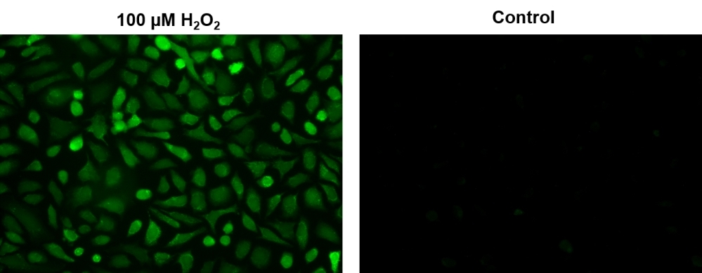
- Monitor the fluorescence increase with a fluorescence plate reader (bottom read mode) at Ex/Em = 490/525 nm (Cutoff = 515 nm) Or image the fluorescence change with a fluorescence microscope using FITC channel.
Images
Citations
Authors: Hu, Can and Qiu, Yan and Guo, Jiajun and Cao, Yuchao and Li, Dairong and Du, Yonghong
Journal: International Journal of Nanomedicine (2023): 6257--6274
Authors: Ishak, Nur Syafiqah Mohamad and Numaguchi, Tomoe and Ikemoto, Kazuto
Journal: ACS Omega (2023)
Authors: Nie, Hongyun and Nie, Maiqian and Diwu, Zhenjun and Wang, Lei and Qiao, Qi and Zhang, Bo and Yang, Xuefu
Journal: Environmental Research (2020): 110159
Authors: Nishide, Masayuki and Nojima, Satoshi and Ito, Daisuke and Takamatsu, Hyota and Koyama, Shohei and Kang, Sujin and Kimura, Tetsuya and Morimoto, Keiko and Hosokawa, Takashi and Hayama, Yoshitomo and others, undefined
Journal: Annals of the Rheumatic Diseases (2017): annrheumdis--2016
Authors: Rong, Lei and Zhang, Chi and Lei, Qi and Hu, Ming-Ming and Feng, Jun and Shu, Hong-Bing and Liu, Yi and Zhang, Xian-Zheng
Journal: Regenerative Biomaterials (2016): rbw022
Authors: Uemura, Takeshi and Watanabe, Kenta and Ishibashi, Misaki and Saiki, Ryotaro and Kuni, Kyoshiro and Nishimura, Kazuhiro and Toida, Toshihiko and Kashiwagi, Keiko and Igarashi, Kazuei
Journal: Biochemical and biophysical research communications (2016): 630--635
Authors: Chen, Cheng-Yu and Lee, Cheng-Cheng and Chen, Hung-Shuan and Yang, Chao-Hsun and Wang, Shu-Ping and Wu, Jyh-Horng and Meng, Menghsiao
Journal: Process Biochemistry (2016): 1486--1495
Authors: Nakaso, Kazuhiro and Tajima, Naoko and Ito, Satoru and Teraoka, Mari and Yamashita, Atsushi and Horikoshi, Yosuke and Kikuchi, Daisuke and Mochida, Shinsuke and Nakashima, Kenji and Matsura, Tatsuya
Journal: PLoS One (2013): e55068
Authors: Ma, He-Ping
Journal: Journal of Biological Chemistry (2011): 32444--32453
Application notes
A Comparison of Fluorescent Red Calcium Indicators for Detecting Intracellular Calcium Mobilization in CHO Cells
A Meta-Analysis of Common Calcium Indicators
A New Red Fluorescent & Robust Screen Quest™ Rhod-4™ Ca2+Indicator for Screening GPCR & Ca2+ Channel Targets
A New Robust No-Wash FLIPR Calcium Assay Kit for Screening GPCR and Calcium Channel Targets