Products
Services
Resources
Selection Guides
About
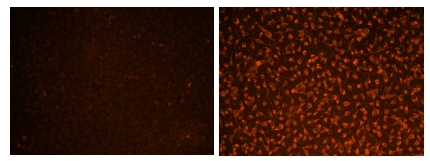
Cell Meter™ No-Wash Live Cell Caspase 3/7 Activity Assay Kit
Red Fluorescence
The activation of caspase 3/7 is important for the initiation of apoptosis. Our Cell Meter™ No-Wash Live Cell Caspase Activity Assay Kits are based on ApoBrite™ V600, our recently developed cell-permeable fluorogenic caspase substrate, the first fluorogenic probe for the direct detection of caspase activities in live cells. ApoBrite V600 consists of three moieties including a). masked fluorophore, b). caspase-selective peptide fragment (DEVD), and c). cell-penetrating moiety. The cell-penetrating moiety carries the probe into live cells. Upon entering live cells the caspase-selective peptide fragment is cleaved by a caspase to release the masked fluorophore. The intensity of recovered fluorescence is directly related to the activity of caspase to be measured. Compared to the existing caspase assays in live cells, ApoBrite™ V600 is much more robust, convenient and accurate. ApoBrite™ V600 releases a fluorophore that has a large Stokes shift, and can be well excited with violet laser that is installed most of new flow cytometers. It does not need a DNA interaction to be fluorescent as reported for NucView reagents. It does not inhibit caspase activity as reported for the FMK peptide probes. Although fluorescent FMK peptide inhibitors of caspases are widely used for detecting caspase activities in live cells, this technology has a few severe limitations: a). FMK caspase inhibitors have high cytotoxicity since FMK peptides bind covalently to active caspases; b). The irreversibly covalent binding of FMK peptides to caspases inhibits caspase activities, causing false positive apoptosis; c). FMK assays have extremely high background, and require intensive washings, resulting in very low through put; d). FMK peptides are not stable in aqueous solutions, and have to be used immediately.
| Catalog | Size | Price | Quantity |
|---|---|---|---|
| 20260 | 200 Tests | Price |
Storage, safety and handling
| H-phrase | H303, H313, H333 |
| Hazard symbol | XN |
| Intended use | Research Use Only (RUO) |
| R-phrase | R20, R21, R22 |
| UNSPSC | 12352200 |
Instrument settings
| Flow cytometer | |
| Excitation | 405 nm |
| Emission | 570 nm |
| Instrument specification(s) | FL3 channel for 7-AAD staining |
| Fluorescence microscope | |
| Excitation | DAPI channel |
| Emission | DAPI channel |
| Recommended plate | Black wall/clear bottom |
| Instrument specification(s) | TRITC channel for 7-AAD staining |
| Fluorescence microplate reader | |
| Excitation | 405 nm |
| Emission | 570 nm |
| Cutoff | 540 nm |
| Recommended plate | Black wall/clear bottom |
| Instrument specification(s) | Bottom read mode |
Contact us
| Telephone | |
| Fax | |
| sales@aatbio.com | |
| International | See distributors |
| Bulk request | Inquire |
| Custom size | Inquire |
| Technical Support | Contact us |
| Request quotation | Request |
| Purchase order | Send to sales@aatbio.com |
| Shipping | Standard overnight for United States, inquire for international |
Page updated on February 1, 2023