logo
AAT Bioquest

Helixyte™ Green Fluorimetric dsDNA Quantitation Kit *High Sensitivity*

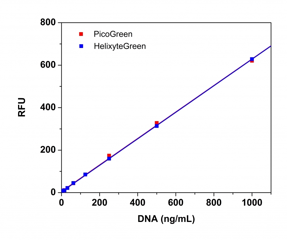
Helixyte™ Green dsDNA Quantitation Assay Kit can be used for selectively detecting as little as 25 pg/ml of dsDNA in the presence of ssDNA, RNA, and free nucleotides. Helixyte™ Green exhibits large fluorescence enhancement upon binding to dsDNA. The assay is linear over three orders of magnitude and has little sequence dependence, allowing you to accurately measure DNA from many sources, including genomic DNA, viral DNA, miniprep DNA, or PCR amplification products. Helixyte™ Green dsDNA Quantitation Assay Kit is a few magnitudes more sensitive than UV absorbance readings. It is specific for dsDNA in the presence of equimolar amounts of RNA. The kit is robust with a mix and read format. It can be used with a bench top fluorometer or a hand-held fluorescence meter (e.g., Qubit fluorometer). This kit is an excellent replacement for Quant-iT™ PicoGreen® dsDNA Assay Kit (Quant-iT™ and PicoGreen® are the trademarks of Invitrogen).
Comparison of dsDNA dose response using the Helixyte Green&trade; (blue) with Invitrogen<sup>TM</sup> Quant-iT&trade; PicoGreen&reg; dsDNA Reagent (red ). dsNDA standards were incubated in cuvettes and measured using varian cary eclipse fluorescence spectrophotometer.&nbsp;
Ordering information
Price
Unit size
Catalog Number17651
Quantity
Add to cart
Additional ordering information
Telephone1-800-990-8053
Fax1-800-609-2943
Emailsales@aatbio.com
InternationalSee distributors
Bulk requestInquire
Custom sizeInquire
ShippingStandard overnight for United States, inquire for international
Request quotation
Spectral properties
Excitation (nm)502
Emission (nm)522
Storage, safety and handling
H-phraseH303, H313, H340
Hazard symbolT
Intended useResearch Use Only (RUO)
R-phraseR20, R21, R68
UNSPSC41116134
Platform

Spectrofluorometer

Excitation490 nm
Emission525 nm
Cutoff515 nm
Components
Example protocol

AT A GLANCE

Protocol Summary
  1. Add 1mL of dsDNA standards or test samples to each cuvette.

  2. Add 1 mL of the Helixyte Green™ working solution.

  3. Incubate at room temperature for 5 to 10 minutes.

  4. Monitor the fluorescence at Ex/Em = 490/525 nm.

Important Note

The following protocol is an example for quantifying dsDNA with Helixyte Green™. Allow all components to warm to room temperature before opening. There is no available data on the mutagenicity or toxicity of Helixyte Green™ dsDNA stain. However, since this reagent binds to nucleic acids, it should be treated as a potential mutagen and handled with appropriate care. Handle the DMSO stock solution with particular caution, as DMSO is known to facilitate the entry of organic molecules into tissues.

PREPARATION OF STOCK SOLUTIONS

Unless otherwise noted, all unused stock solutions should be divided into single-use aliquots and stored at -20 °C after preparation. Avoid repeated freeze-thaw cycles

Assay Buffer (1X)
  1. Prepare a 1X Assay buffer by diluting the concentrated buffer 20-fold with sterile, distilled, DNase-free water.

PREPARATION OF STANDARD SOLUTIONS

For convenience, use the Serial Dilution Planner:
https://www.aatbio.com/tools/serial-dilution/17651

dsDNA standard
For high range standard curve:Add 30 µL of 100 µg/mL dsDNA stock solution (Component C) to 1.47 mL of 1X Assay buffer to have 2000 ng/mL dsDNA solution, and then perform 1:2 and 1:10 serial dilutions to get 1000, 100, 10, 1 and 0 ng/mL.For low range standard curve:Add 40 µL of 2 µg/mL dsDNA stock solution to 1.56 mL of 1X Assay buffer to have 50 ng/mL dsDNA solution, and then perform 1:2 and 1:10 serial dilutions to get 25, 2.5, 0.25, 0.025 and 0 ng/mL.

PREPARATION OF WORKING SOLUTION

Helixyte Green™ Working Solution
  1. To prepare the Helixyte Green™ working solution, make a 200-fold dilution of the concentrated DMSO solution in 1X assay buffer. For example, to prepare enough working solution to assay 10 samples in a final volume of 2 mL each, add 50 μL of Helixyte Green™ (Component A) into 10 mL of Assay Buffer (Component B). Protect the working solution from light by covering it with foil or placing it in a dark location.

    Note: We recommend using a plastic container to prepare this solution, as the dye can adhere to glass surfaces. For optimal performance, use the solution within a few hours of preparation.

SAMPLE EXPERIMENTAL PROTOCOL

  1. Add 1 mL of Helixyte Green™ working solution to each cuvette containing 1 mL of the dsDNA standard, blank control, and test samples. This will bring the total volume of the dsDNA assay in each cuvette to 2 mL.

  2. Incubate the reaction at room temperature for 5-10 minutes, keeping it protected from light.

  3. Monitor the fluorescence increase with a spectrofluorometer at Ex/Em = 490/525 nm, Cutoff = 515 nm.

    Note: To minimize photobleaching effects, keep the time for fluorescence measurement constant for all samples.

Spectrum
Citations
View all 1 citations: Citation Explorer
A cellular assay to determine the fusion capacity of MFN2 variants linked to Charcot-Marie-Tooth type 2A
Authors: Barsa, Chloe and Perrin, Julian and David, Claudine and Mourier, Arnaud and Rojo, Manuel
Journal: bioRxiv (2024): 2024--03
References
View all 31 references: Citation Explorer
Inhibitors of Streptococcus pneumoniae surface endonuclease EndA discovered by high-throughput screening using a PicoGreen fluorescence assay
Authors: Peterson EJ, Kireev D, Moon AF, Midon M, Janzen WP, Pingoud A, Pedersen LC, Singleton SF.
Journal: J Biomol Screen (2013): 247
Validation of a PicoGreen-based DNA quantification integrated in an RNA extraction method for two-dimensional and three-dimensional cell cultures
Authors: Chen Y, Sonnaert M, Roberts SJ, Luyten FP, Schrooten J.
Journal: Tissue Eng Part C Methods (2012): 444
Characterization of PicoGreen interaction with dsDNA and the origin of its fluorescence enhancement upon binding
Authors: Dragan AI, Casas-Finet JR, Bishop ES, Strouse RJ, Schenerman MA, Geddes CD.
Journal: Biophys J (2010): 3010
Comparison of SYBR Green I-, PicoGreen-, and [3H]-hypoxanthine-based assays for in vitro antimalarial screening of plants from Nigerian ethnomedicine
Authors: Abiodun OO, Gbotosho GO, Ajaiyeoba EO, Happi CT, Hofer S, Wittlin S, Sowunmi A, Brun R, Oduola AM.
Journal: Parasitol Res (2010): 933
Metal-enhanced PicoGreen fluorescence: application to fast and ultra-sensitive pg/ml DNA quantitation
Authors: Dragan AI, Bishop ES, Casas-Finet JR, Strouse RJ, Schenerman MA, Geddes CD.
Journal: J Immunol Methods (2010): 95