PhosphoWorks™ Luminometric ATP Assay Kit *
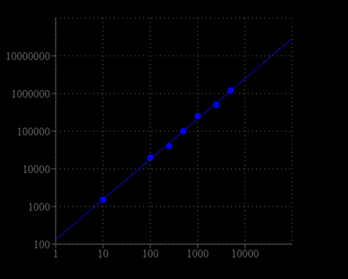
Maximized Luminescence*
![CHO-K1 cell number was measured with PhosphoWorks™ Luminescence ATP Assay Kit on a 96-well white plate using a NOVOstar plate reader (BMG Labtech). The integration time was 1 sec.](/_next/image?url=https%3A%2F%2Fimages.aatbio.com%2Fproducts%2Ffigures-and-data%2Fphosphoworks-luminometric-atp-assay-kit-bright-glow%2Fgraph-for-phosphoworks-luminometric-atp-assay-kit-maximized-luminescence_tDsM0.webp&w=640&q=75)
![CHO-K1 cell number was measured with PhosphoWorks™ Luminescence ATP Assay Kit on a 96-well white plate using a NOVOstar plate reader (BMG Labtech). The integration time was 1 sec.](/_next/image?url=https%3A%2F%2Fimages.aatbio.com%2Fproducts%2Ffigures-and-data%2Fphosphoworks-luminometric-atp-assay-kit-bright-glow%2Fgraph-for-phosphoworks-luminometric-atp-assay-kit-maximized-luminescence_tDsM0.webp&w=128&q=25)
![Impact of host-cell metabolism inhibition on ATP levels and τ2-NAD(P)H. Cellular ATP levels under glucose starvation and inhibition of oxidative phosphorylation by antimycin A. *ATP was measured using a luminometric ATP assay kit (ABD Bioquest, Sunnyvale, CA) and a microplate reader (Tecan Infinite 200 PRO, Maenedorf, Switzerland). HEp-2 cells were grown in 96-well plates (2000 cells/ well) and treated with the metabolic inhibitors as described above. ATP assays were performed according to the manufacturer's instructions. Source: Graph from <strong>Fluorescence Lifetime Imaging Unravels C. trachomatis Metabolism and Its Crosstalk with the Host Cell</strong> by Márta Szaszák, et al., <em>PLOS ONE</em>, July 2011. ](/_next/image?url=https%3A%2F%2Fimages.aatbio.com%2Fproducts%2Ffigures-and-data%2Fphosphoworks-luminometric-atp-assay-kit-bright-glow%2Ffigure-for-phosphoworks-luminometric-atp-assay-kit-maximized-luminescence_KByOL.jpg&w=128&q=25)
![Optimization of the indirect glycolate cytotoxicity. (A) A collection of eight CHO cell lines were cultured in 384-well plates and assayed for the glycolate toxicity. (B–D) Optimization of assay parameters was carried out in three aspects: seeding CHO cells at different density (B), adding glycolate at a range of concentrations (C), and varying the incubation period (D). Signal-to-basal ratio was evaluated for each parameter. (E) Partial reduction of the indirect glycolate cytotoxicity in the cell-based assay by 2-hydroxy-3-butynoic acid (2H3BA) c, a known GO inhibitor. ATP content cell viability assay using an assay kit (21610, AAT Bioquest). Source: <b>High throughput cell-based assay for identification of glycolate oxidase inhibitors as a potential treatment for Primary Hyperoxaluria Type 1</b> by Wang M, Xu M, Long Y, Fargue S, Southall N, Hu X, McKew JC, Danpure CJ, Zheng W. High. <em>Sci Rep.</em>, Sep. 2016.](/_next/image?url=https%3A%2F%2Fimages.aatbio.com%2Fproducts%2Ffigures-and-data%2Fphosphoworks-luminometric-atp-assay-kit-bright-glow%2Ffigure-for-phosphoworks-luminometric-atp-assay-kit-maximized-luminescence_GG5C3.jpeg&w=128&q=25)
AT A GLANCE
Protocol summary
- Prepare cells (samples) with test compounds (100 µL/96-well plate or 25 µL/384-well plate)
- Add equal volume of ATP working solution (100 µL/96-well plate or 25 µL/384-well plate)
- Incubate at room temperature for 10 - 20 minutes
- Monitor the luminescence intensity
Important notes
To achieve the best results, it’s strongly recommended to use the white plates. Thaw all the kits components at room temperature before starting the experiment.
PREPARATION OF WORKING SOLUTION
1. Transfer the whole content of Reaction Buffer (Component C, 10 mL) into ATP Sensor (Component B) and mix well.
2. Add 20 µL of ATP Monitoring Enzyme (Component A) into the bottle of Component B+C and mix well to make ATP working solution. Note: Avoid potential ATP contamination from exogenous biological sources.
For guidelines on cell sample preparation, please visit
https://www.aatbio.com/resources/guides/cell-sample-preparation.html
SAMPLE EXPERIMENTAL PROTOCOL
Run ATP assay:
- Treat cells (or samples) with test compounds by adding 10 µL of 10X compounds for a 96-well plate or 5 µL of 5X compounds for a 384-well plate in desired compound buffer. For blank wells (medium without the cells), add the corresponding amount of compound buffer.
- Incubate the cell plate in a 37°C, 5% CO2 incubator for a desired period of time, such as 24, 48 or 96 hours.
- Add 100 µL (96-well plate) or 25 µL (384-well plate) of ATP working solution into each well.
- Incubate at room temperature for 10 - 20 minutes.
- Monitor luminescence intensity with a standard luminometer.
Generate a standard ATP calibration curve:
An ATP standard curve should be generated together with the above assay if the absolute amount of ATP in samples needs to be calculated.
- Make a series of dilutions of ATP in PBS buffer with 0.1% BSA by including a sample without ATP (as a control) for measuring background luminescence. Note: Typically ATP concentrations from 1 nM to 10 µM are appropriate.
- Add the same amount of the diluted ATP solution into an empty plate (100 µL for a 96-well plate or 25 µL for a 384-well plate).
- Add 100 µL/well (96-well plate) or 25 µL/well (384-well plate) of ATP working solution.
- Incubate the reaction mixture at room temperature for 10 to 20 minutes.
- Monitor the luminescence intensity with a standard luminometer.
- Generate the ATP standard curve.
Authors: Zhao, Ziyi and Wang, Jiandong and Kong, Weimin and Newton, Meredith A and Burkett, Wesley C and Sun, Wenchuan and Buckingham, Lindsey and O’Donnell, Jillian and Suo, Hongyan and Deng, Boer and others,
Journal: Biomolecules (2024): 601
Authors: Qiu, Bin and Zhong, Zhaohui and Dou, Longyu and Xu, Yuxue and Zou, Yi and Weldon, Korri and Wang, Jun and Zhang, Lingling and Liu, Ming and Williams, Kent E and others,
Journal: Cell \& Bioscience (2024): 1
Authors: Li, Yao and Xia, Yuchen and Cheng, Xiaoming and Kleiner, David E and Hewitt, Stephen M and Sproch, Julia and Li, Tong and Zhuang, Hui and Liang, T Jake
Journal: Viruses (2019): 386
Authors: Chen, Ya-Hui and Chen, Yi-Chun and Liu, Chin-San and Hsieh, Ming-Chia
Journal: Journal of Diabetes Research (2016)
Authors: Zhang, Lu and Han, Jianjun and Jackson, Am and a L , undefined and Clark, Leslie N and Kilgore, Joshua and Guo, Hui and Livingston, Nick and Batchelor, Kenneth and Yin, Yajie and Gilliam, Timothy P and others, undefined
Journal: Journal of Hematology & Oncology (2016): 91