Endoplasmic Reticulum (ER)
The endoplasmic reticulum (ER) is an organelle present in most eukaryotic cells. It primarily synthesizes and transports cellular products such as proteins, lipids, and hormones. Structurally, the ER comprises an interconnected network of flattened, membrane-enclosed sacs and tubules extending from the nuclear membrane throughout the cytoplasm. These sac-like structures and tubules are held together and supported by the cytoskeleton.
The ER complex comprises two subunits - rough ER and smooth SER. The surface of the rough ER is coated with protein-manufacturing ribosomes. Once synthesized, membrane-bound transport vesicles shuttle these proteins to the Golgi apparatus. The smooth ER lacks ribosomes and is responsible for synthesizing lipids, phospholipids, and steroids. Secondary responsibilities of the smooth ER include carbohydrate and steroid metabolism, detoxification, and modulation of calcium ions.
Endoplasmic Reticulum Staining Kits for Live Cells
ER Tracer™ dyes are membrane-permeant stains considerably selective for the endoplasmic reticulum in living cells. As the key component in our Cell Navigator™ Live Cell Endoplasmic Reticulum Staining Kits, ER Tracer™ dyes can be multiplexed with other fluorescent proteins or probes in live cell multiparametric studies or after fixation for colocalization studies. However, ER Tracer™ stains may not selectively bind to ER for certain cell lines.
Key Features of Cell Navigator™ Live Cell ER Staining Kits:
- Highly selective for ER over other cellular compartments (e.g., lysosomes and mitochondria)
- ER staining well-retained after cell fixation
- Available in blue, green, and red fluorescence to facilitate multiplexing with other fluorescent probes
- Suitable for proliferating and non-proliferating cells
- Kits include a robust staining protocol and sufficient materials for 100 tests

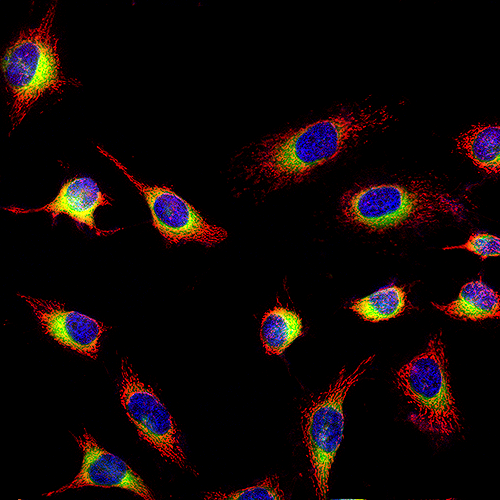

Table 1. Cell Navigator® ER Staining Kits for imaging the endoplasmic reticulum of live cells.
| Kit ▲ ▼ | Ex (nm) ▲ ▼ | Em (nm) ▲ ▼ | Filter Set ▲ ▼ | Unit Size ▲ ▼ | Cat No. ▲ ▼ |
| Cell Navigator® Live Cell ER Staining Kit *Blue Fluorescence* | 344 | 457 | DAPI filter set | 100 Tests | 22634 |
| Cell Navigator® Live Cell ER Staining Kit *Green Fluorescence* | 503 | 511 | FITC filter set | 100 Tests | 22635 |
| Cell Navigator® Live Cell ER Staining Kit *Red Fluorescence* | 588 | 620 | Texas Red filter set | 100 Tests | 22636 |