logo
AAT Bioquest

Buccutite™ Rapid APC-Cy5.5 Tandem Antibody Labeling Kit *Production Scale Optimized for Labeling 1 mg Antibody Per Reaction*

Human peripheral blood lymphocytes were stained with APC-Cy5.5 anti-Human CD4 (clone SK3, mouse IgG1, κ) conjugate prepared with APC-Cy5.5 Tandem (Cat# 2622). APC-Cy5.5 anti-human CD4 *SK3* conjugates were prepared using the Buccutite™ Rapid APC-Cy5.5 Tandem Antibody Labeling Kit *Production Scale* (Cat# 5414).
Ordering information
Price
Catalog Number
Unit Size
Quantity
Add to cart
Additional ordering information
Telephone1-800-990-8053
Fax1-800-609-2943
Emailsales@aatbio.com
InternationalSee distributors
Bulk requestInquire
Custom sizeInquire
ShippingStandard overnight for United States, inquire for international
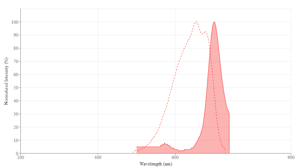
Request quotation
Spectral properties
Extinction coefficient (cm -1 M -1)700000
Excitation (nm)651
Emission (nm)700
Storage, safety and handling
H-phraseH303, H313, H333
Hazard symbolXN
Intended useResearch Use Only (RUO)
R-phraseR20, R21, R22
UNSPSC12171501
Related products
Buccutite™ Rapid PE Antibody Labeling Kit *Microscale Optimized for Labeling 100 ug Antibody Per Reaction*
Buccutite™ Rapid APC Antibody Labeling Kit *Microscale Optimized for Labeling 100 ug Antibody Per Reaction*
Buccutite™ Peroxidase (HRP) Antibody Conjugation Kit *Optimized for Labeling 100 ug Protein*
Buccutite™ Rapid Protein Crosslinking Kit *Microscale Optimized for Crosslinking 100 ug Antibody Per Reaction*
Buccutite™ Rapid PE Antibody Labeling Kit *Microscale Optimized for Labeling 25 ug Antibody Per Reaction*
Buccutite™ Rapid APC Antibody Labeling Kit *Microscale Optimized for Labeling 25 ug Antibody Per Reaction*
Buccutite™ Rapid PE-Cy7 Tandem Antibody Labeling Kit *Microscale Optimized for Labeling 100 ug Antibody Per Reaction*
Buccutite™ Rapid PE-Texas Red Tandem Antibody Labeling Kit *Microscale Optimized for Labeling 100 ug Antibody Per Reaction*
Buccutite™ Rapid PE-Cy5 Tandem Antibody Labeling Kit *Microscale Optimized for Labeling 100 ug Antibody Per Reaction*
Buccutite™ Rapid APC-iFluor® 700 Tandem Antibody Labeling Kit *Microscale Optimized for Labeling 100 ug Antibody Per Reaction*
Buccutite™ Rapid APC-Cy7 Tandem Antibody Labeling Kit *Microscale Optimized for Labeling 100 ug Antibody Per Reaction*
Buccutite™ Rapid PerCP Antibody Labeling Kit *Microscale Optimized for Labeling 100 ug Antibody Per Reaction*
Buccutite™ Rapid PE-Cy5 Tandem Antibody Labeling Kit *Microscale Optimized for Labeling 25 ug Antibody Per Reaction*
Buccutite™ Rapid PE-Cy7 Tandem Antibody Labeling Kit *Microscale Optimized for Labeling 25 ug Antibody Per Reaction*
Buccutite™ Rapid PE-Texas Red Tandem Antibody Labeling Kit *Microscale Optimized for Labeling 25 ug Antibody Per Reaction*
Buccutite™ Rapid APC-iFluor® 700 Tandem Antibody Labeling Kit *Microscale Optimized for Labeling 25 ug Antibody Per Reaction*
Buccutite™ Rapid APC-Cy7 Tandem Antibody Labeling Kit *Microscale Optimized for Labeling 25 ug Antibody Per Reaction*
Buccutite™ Rapid PerCP Antibody Labeling Kit *Microscale Optimized for Labeling 25 ug Antibody Per Reaction*
Buccutite™ Peroxidase (HRP) Antibody Conjugation Kit *Optimized for Labeling 25 ug Protein*
Buccutite™ Peroxidase (HRP) Antibody Conjugation Kit *Optimized for Labeling 1 mg Protein*
Buccutite™ MTA, NHS ester
Buccutite™ FOL scavenger
Buccutite™ MTA scavenger
Buccutite™ MTA-Dye 650
Buccutite™ FOL-Dye 650
Buccutite™ ALP (Alkaline Phosphatase) Antibody Conjugation Kit *Optimized for Labeling 25 ug Protein*
Buccutite™ ALP (Alkaline Phosphatase) Antibody Conjugation Kit *Optimized for Labeling 100 ug Protein*
Buccutite™ ALP (Alkaline Phosphatase) Antibody Conjugation Kit *Optimized for Labeling 1 mg Protein*
Buccutite™ Poly-HRP Antibody Conjugation Kit
Buccutite™ FOL, maleimide [FOLM]
Buccutite™ MTA, maleimide [MTAM]
Buccutite™ FOL, NHS ester
Buccutite™ Rapid APC-iFluor® 750 Tandem Antibody Labeling Kit *Microscale Optimized for Labeling 25 ug Antibody Per Reaction*
Buccutite™ Rapid trFluor™ D2 Acceptor Antibody Labeling Kit *Microscale Optimized for Labeling 100 ug Antibody Per Reaction*
Buccutite™ Streptavidin Antibody Conjugation Kit *Optimized for Labeling 25 ug Protein*
Buccutite™ Streptavidin Antibody Conjugation Kit *Optimized for Labeling 100 ug Protein*
Buccutite™ Streptavidin Antibody Conjugation Kit *Optimized for Labeling 1 mg Protein*
Buccutite™ Rapid Oligo Antibody Conjugation Kit *Optimized for Labeling 100 ug Protein*
Buccutite™ Rapid PE Antibody Labeling Kit *Production Scale Optimized for Labeling 1 mg Antibody Per Reaction*
Buccutite™ Rapid Crosslinked APC Antibody Labeling Kit *Production Scale Optimized for Labeling 1 mg Antibody Per Reaction*
Buccutite™ Rapid PerCP Antibody Labeling Kit *Production Scale Optimized for Labeling 1 mg Antibody Per Reaction*
Buccutite™ Rapid PE-Cy7 Tandem Antibody Labeling Kit *Production Scale Optimized for Labeling 1 mg Antibody Per Reaction*
Buccutite™ Rapid PE-Texas Red Tandem Antibody Labeling Kit *Production Scale Optimized for Labeling 1 mg Antibody Per Reaction*
Buccutite™ Rapid PE-Cy5 Tandem Antibody Labeling Kit *Production Scale Optimized for Labeling 1 mg Antibody Per Reaction*
Buccutite™ Rapid APC-iFluor® 700 Tandem Antibody Labeling Kit *Production Scale Optimized for Labeling 1 mg Antibody Per Reaction*
Buccutite™ Rapid APC-Cy7 Tandem Antibody Labeling Kit *Production Scale Optimized for Labeling 1 mg Antibody Per Reaction*
Show More (36)

OverviewpdfSDSpdfProtocol


Extinction coefficient (cm -1 M -1)
700000
Excitation (nm)
651
Emission (nm)
700
Buccutite™ Rapid APC-Cy5.5 Tandem Antibody Labeling Kits, designed for large-scale production, provide a streamlined approach for labeling antibodies with PE, APC, PerCP, and iFluor® tandem dyes. Compared to conventional protein-protein conjugation methods like the SMCC crosslinking technique, Buccutite™ conjugation is simple and more robust. Using a two-step mixing protocol, researchers can directly conjugate APC-Cy5.5 to any antibody or protein in less than 2 hours. Each Buccutite™ kit includes all the essential components for two labeling reactions and features a user-friendly, pre-packed spin column for maximum conjugate yield. Each Buccutite™ FOL-Activated APC-Cy5.5 vial provided in this kit is precisely formulated to label 1 mg of purified protein or antibody. Before labeling, it's important to remove stabilizing proteins like BSA from the sample and avoid using amine-rich buffers like Tris, which might disrupt the labeling process. Allophycocyanin-cyanine 5.5 (APC-Cy5.5) is an intensely bright, red fluorescent tandem fluorophore with an excitation and emission maxima of ~651 nm and ~700 nm, respectively. Given its intense brightness, APC-Cy5.5 is recommended for pairing with low-abundance targets to minimize spillover and compensation. APC-Cy5.5 conjugates are well-suited for flow cytometry, spectral flow cytometry, and other immunoassays requiring high sensitivity but not photostability. With Buccuitte™ Rapid Antibody Labeling kits, researchers can directly label primary antibodies, eliminating the need for secondary antibodies and enhancing panel-building flexibility.

Components


Example protocol


AT A GLANCE

Key Parameters to Achieve Best Performance
  1. 1.0 mg Antibody (MW ~150 kDa)

  2. Antibody concentration: 2.0 mg/mL

  3. Antibody volume: 500 µL

PREPARATION OF WORKING SOLUTION

Important

Before opening the vials, warm all components and briefly centrifuge. Immediately prepare necessary solutions before starting conjugation. This protocol is a recommendation.

Prepare Antibody Solution
  1. Prepare a 500 µL antibody solution in PBS with a concentration of 2 mg/mL.

    Note: The protein should be dissolved in 1X phosphate-buffered saline (PBS), pH 7.2 - 7.4. If the protein is dissolved in buffers containing primary amines, like Tris and/or glycine, it must be dialyzed against 1X PBS, pH 7.2 - 7.4, or use Amicon Ultra0.5, Ultracel-10 Membrane, 10 kDa (Cat No. UFC501008 from Millipore) to remove free amines or ammonium salts (such as ammonium sulfate and ammonium acetate) that are widely used for protein precipitation.

    Note: Impure antibodies or antibodies stabilized with bovine serum albumin (BSA) or gelatin will not be labeled well.

Prepare Buccutite™ MTA Solution
  1. Warm up a vial of Buccutite™ MTA (Component B) to room temperature.

  2. Add 5 µL of DMSO (not provided) to the vial of Buccutite™ MTA (Component B), and mix well by pipetting.

SAMPLE EXPERIMENTAL PROTOCOL

Run Antibody-Buccutite™ MTA Reaction
  1. Add 25 µL of Reaction Buffer (Component C) to the antibody solution.

  2. Transfer 5 µL of the reconstituted Buccutite™ MTA DMSO solution into the vial of antibody solution, and mix well by pipetting.

  3. Rotate the reaction mixture at room temperature for 1 hour, then purify using a desalting column.

Purify Antibody-Buccutite™ MTA Solution with Desalting Column
  1. Invert the provided spin column (Component D) several times to re-suspend the settled gel and remove any bubbles.

  2. Snap off the tip and place the column in a washing tube (2 mL, not provided). Remove the cap to allow the excess packing buffer to drain by gravity to the top of the gel bed.

    Note: If the column does not begin to flow, push the cap back into the column and remove it again to start the flow. Discard the drained buffer, and then place the column back into the Washing Tube. 

  3. Centrifuge at 1000 x g for 2 minutes in a swinging bucket centrifuge to remove the packing buffer. Then discard the buffer. Refer to the 'Centrifugation Notes' section below for instructions.

  4. Apply 1-2 mL 1X PBS (pH 7.2-7.4) to the column. After each application of PBS, let the buffer drain out by gravity, or centrifuge the column for 2 minutes to remove the buffer. Discard the buffer from the collection tube. Repeat this process for 3-4 times.

  5. Centrifuge at 1000 x g for 2 minutes in a swinging bucket centrifuge to remove the packing buffer. Then discard the buffer. Refer to the 'Centrifugation Notes' section below for instructions.

  6. Place the column into a clean collecting tube (1.5 mL, not provided). Then, take the antibody-Buccutite™ MTA solution from step 3 of the "Run Antibody-Buccutite™ MTA Reaction" section and load it carefully and directly into the center of the column.

  7. After loading the sample, add 40 μL of 1X PBS (pH 7.2-7.4), centrifuge the column for 2 minutes at 1,000 x g, and collect the solution that contains the desired antibody-Buccutite™ MTA solution.

Run Antibody-APC-Cy5.5 Conjugation Reaction
  1. Warm up a vial of Buccutite™ FOL-Activated APC-Cy5.5 (Component A) to room temperature.

    Note: Each vial of Buccutite™ FOL-Activated APC-Cy5.5 contains an optimized amount of dye to label 1 mg of IgG (MW ~150 kDa) at 2 mg/mL in PBS, the kit can also be used to label other proteins (>10 kDa).

  2. Make a Buccutite™ FOL-Activated APC-Cy5.5 solution by adding 130 µL of ddH2O into the vial of Buccutite™ FOL-Activated APC-Cy5.5 (Component A), and mix well by pipetting or vortexing.

  3. Add the purified Antibody-Buccutite™ MTA solution directly into the vial of Buccutite™ FOL-Activated APC-Cy5.5 solution. Rotate the mixture for 1-2 hours at room temperature.

  4. The antibody-APC-Cy5.5 conjugate is now ready for immediate use or can be stored at 4°C.

Purification with Size Exclusion Chromatography Recommended
  1. For optimal performance, it is recommended to purify the antibody-APC-Cy5.5 conjugate using size exclusion chromatography (SEC). The following SEC columns are suitable for this purpose: Superdex 200 Increase 100/300 GL (Cytiva) and ENrich™ SEC 650 10 x 300 Column (Bio-Rad).

Spectrum


Open in Advanced Spectrum Viewer
spectrum

Spectral properties

Extinction coefficient (cm -1 M -1)700000
Excitation (nm)651
Emission (nm)700

Images


References


View all 34 references: Citation Explorer
Single-Cell Sorting of Immunophenotyped Mesenchymal Stem Cells from Human Exfoliated Deciduous Teeth.
Authors: Gupta, Ayona and Mukhopadhyay, Risani and Khandelwal, Himanshi and Nala, Narendra and Chakraborty, Uttara
Journal: Journal of visualized experiments : JoVE (2023)
Recombinant thrombomodulin attenuates hyper-inflammation and glycocalyx damage in a murine model of Streptococcus pneumoniae-induced sepsis.
Authors: Watanabe, Eizo and Akamatsu, Toshinobu and Ohmori, Masaaki and Kato, Mayu and Takeuchi, Noriko and Ishiwada, Naruhiko and Nishimura, Rintaro and Hishiki, Haruka and Fujimura, Lisa and Ito, Chizuru and Hatano, Masahiko
Journal: Cytokine (2022): 155723
Routine flow cytometry approach for the evaluation of solid tumor neoplasms and immune cells in minimally invasive samples.
Authors: Quirós-Caso, Covadonga and Arias Fernández, Tamara and Fonseca-Mourelle, Ariana and Torres, Héctor and Fernández, Luis and Moreno-Rodríguez, Maria and Ariza-Prota, Miguel Ángel and López-González, Francisco Julián and Carvajal-Álvarez, Miguel and Alonso-Álvarez, Sara and Moro-García, Marco Antonio and Colado, Enrique
Journal: Cytometry. Part B, Clinical cytometry (2022)
The hernia sac-A suitable source for obtaining mesenchymal stem cells.
Authors: Lin, Alpha Dian-Yu and Tung, Min-Che and Lu, Chin-Heng
Journal: Surgery open science (2021): 40-44
The ISCCA flow protocol for the monitoring of anti-CD20 therapies in autoimmune disorders.
Authors: Gatti, Arianna and Buccisano, Francesco and Scupoli, Maria T and Brando, Bruno
Journal: Cytometry. Part B, Clinical cytometry (2021): 194-205
Identification of circulating cells interacted with integrin α4β1 ligand peptides REDV or HGGVRLY.
Authors: Hsu, Yu-I and Mahara, Atsushi and Yamaoka, Tetsuji
Journal: Peptides (2021): 170470
Integrated Imaging Methodology Detects Claudin-1 Expression in Premalignant Nonpolypoid and Polypoid Colonic Epithelium in Mice.
Authors: Wang, Fa and Duan, Xiyu and Chen, Jing and Gao, Zhenghong and Zhou, Juan and Wu, Xiaoli and Chang, Tse-Shao and Lee, Miki and Li, Gaoming and Nusrat, Asma and Kuick, Rork and Appelman, Henry D and Wang, Thomas D
Journal: Clinical and translational gastroenterology (2020): e00089
Comparison of minimal residual disease detection in multiple myeloma by SRL 8-color single-tube and EuroFlow 8-color 2-tube multiparameter flow cytometry.
Authors: Takamatsu, Hiroyuki and Yoroidaka, Takeshi and Fujisawa, Momoko and Kobori, Kazuya and Hanawa, Masako and Yamashita, Takeshi and Murata, Ryoichi and Ueda, Mikio and Nakao, Shinji and Matsue, Kosei
Journal: International journal of hematology (2019): 377-381
Detection of colonic neoplasia in vivo using near-infrared-labeled peptide targeting cMet.
Authors: Wu, Xiaoli and Zhou, Juan and Wang, Fa and Meng, Xiaoqing and Chen, Jing and Chang, Tse-Shao and Lee, Miki and Li, Gaoming and Li, Xue and Appelman, Henry D and Kuick, Rork and Wang, Thomas D
Journal: Scientific reports (2019): 17917
Initial flow cytometric evaluation of the Clearllab lymphoid screen.
Authors: Hedley, B D and Cheng, G and Luider, J and Kern, W and Lozanski, G and Chin-Yee, I and Lowes, L E and Keeney, M and Careaga, D and Magari, R and Tejidor, L
Journal: Cytometry. Part B, Clinical cytometry (2018): 707-713