Buccutite™ Rapid PE-Cy7 Tandem Antibody Labeling Kit *Production Scale Optimized for Labeling 1 mg Antibody Per Reaction*
| Price | |
| Catalog Number | |
| Unit Size | |
| Quantity |
| Telephone | 1-800-990-8053 |
| Fax | 1-800-609-2943 |
| sales@aatbio.com | |
| International | See distributors |
| Bulk request | Inquire |
| Custom size | Inquire |
| Shipping | Standard overnight for United States, inquire for international |
| Extinction coefficient (cm -1 M -1) | 1960000 |
| Excitation (nm) | 565 |
| Emission (nm) | 778 |
| H-phrase | H303, H313, H333 |
| Hazard symbol | XN |
| Intended use | Research Use Only (RUO) |
| R-phrase | R20, R21, R22 |
| UNSPSC | 12171501 |
| Overview |
Extinction coefficient (cm -1 M -1) 1960000 | Excitation (nm) 565 | Emission (nm) 778 |
Components
Example protocol
AT A GLANCE
1.0 mg Antibody (MW ~150 kDa)
Antibody concentration: 2.0 mg/mL
Antibody volume: 500 µL
PREPARATION OF WORKING SOLUTION
Before opening the vials, warm all components and briefly centrifuge. Immediately prepare necessary solutions before starting conjugation. This protocol is a recommendation.
Prepare a 500 µL antibody solution in PBS with a concentration of 2 mg/mL.
Note: The protein should be dissolved in 1X phosphate-buffered saline (PBS), pH 7.2 - 7.4. If the protein is dissolved in buffers containing primary amines, like Tris and/or glycine, it must be dialyzed against 1X PBS, pH 7.2 - 7.4, or use Amicon Ultra0.5, Ultracel-10 Membrane, 10 kDa (Cat No. UFC501008 from Millipore) to remove free amines or ammonium salts (such as ammonium sulfate and ammonium acetate) that are widely used for protein precipitation.
Note: Impure antibodies or antibodies stabilized with bovine serum albumin (BSA) or gelatin will not be labeled well.
Warm up a vial of Buccutite™ MTA (Component B) to room temperature.
Add 5 µL of DMSO (not provided) to the vial of Buccutite™ MTA (Component B), and mix well by pipetting.
SAMPLE EXPERIMENTAL PROTOCOL
Add 25 µL of Reaction Buffer (Component C) to the antibody solution.
Transfer 5 µL of the reconstituted Buccutite™ MTA DMSO solution into the vial of antibody solution, and mix well by pipetting.
Rotate the reaction mixture at room temperature for 1 hour, then purify using a desalting column.
Invert the provided spin column (Component D) several times to re-suspend the settled gel and remove any bubbles.
Snap off the tip and place the column in a washing tube (2 mL, not provided). Remove the cap to allow the excess packing buffer to drain by gravity to the top of the gel bed.
Note: If the column does not begin to flow, push the cap back into the column and remove it again to start the flow. Discard the drained buffer, and then place the column back into the Washing Tube.
Centrifuge at 1000 x g for 2 minutes in a swinging bucket centrifuge to remove the packing buffer. Then discard the buffer. Refer to the 'Centrifugation Notes' section below for instructions.
Apply 1-2 mL 1X PBS (pH 7.2-7.4) to the column. After each application of PBS, let the buffer drain out by gravity, or centrifuge the column for 2 minutes to remove the buffer. Discard the buffer from the collection tube. Repeat this process for 3-4 times.
Centrifuge at 1000 x g for 2 minutes in a swinging bucket centrifuge to remove the packing buffer. Then discard the buffer. Refer to the 'Centrifugation Notes' section below for instructions.
Place the column into a clean collecting tube (1.5 mL, not provided). Then, take the antibody-Buccutite™ MTA solution from step 3 of the "Run Antibody-Buccutite™ MTA Reaction" section and load it carefully and directly into the center of the column.
After loading the sample, add 40 μL of 1X PBS (pH 7.2-7.4), centrifuge the column for 2 minutes at 1,000 x g, and collect the solution that contains the desired antibody-Buccutite™ MTA solution.
Warm up a vial of Buccutite™ FOL-Activated PE-Cy7 (Component A) to room temperature.
Note: Each vial of Buccutite™ FOL-Activated PE-Cy7 contains an optimized amount of dye to label 1 mg of IgG (MW ~150 kDa) at 2 mg/mL in PBS, the kit can also be used to label other proteins (>10 kDa).
Make a Buccutite™ FOL-Activated PE-Cy7 solution by adding 250 µL of ddH2O into the vial of Buccutite™ FOL-Activated PE-Cy7 (Component A), and mix well by pipetting or vortexing.
Add the purified Antibody-Buccutite™ MTA solution directly into the vial of Buccutite™ FOL-Activated PE-Cy7 solution. Rotate the mixture for 1-2 hours at room temperature.
The antibody-PE-Cy7 conjugate is now ready for immediate use or can be stored at 4°C.
For optimal performance, it is recommended to purify the antibody-PE-Cy7 conjugate using size exclusion chromatography (SEC). The following SEC columns are suitable for this purpose: Superdex 200 Increase 100/300 GL (Cytiva) and ENrich™ SEC 650 10 x 300 Column (Bio-Rad).
Spectrum
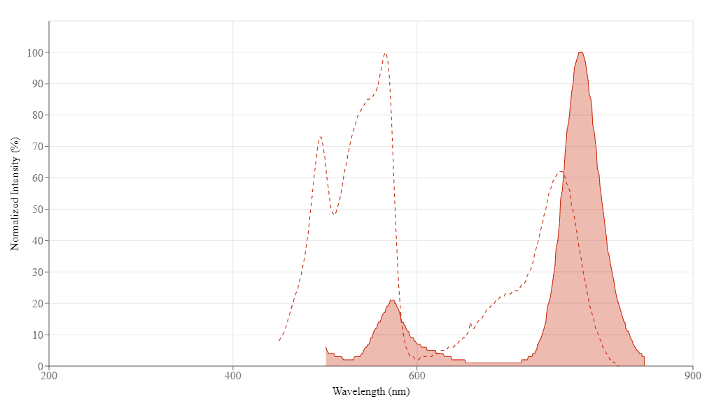
Spectral properties
| Extinction coefficient (cm -1 M -1) | 1960000 |
| Excitation (nm) | 565 |
| Emission (nm) | 778 |
Product Family
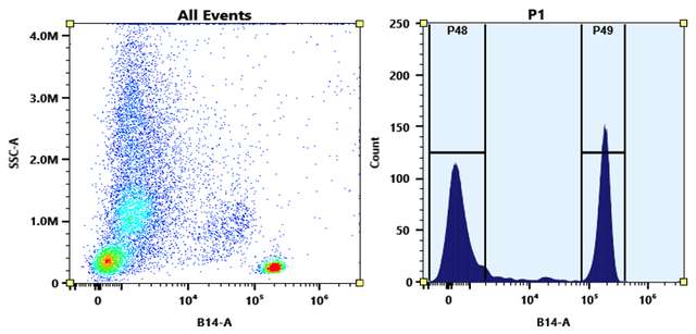
Images

References
Authors: Waeckel, Louis and Khenine, Hana and Berger, Anne-Emmanuelle and Lambert, Claude
Journal: Cytometry. Part A : the journal of the International Society for Analytical Cytology (2023)
Authors: Watanabe, Eizo and Akamatsu, Toshinobu and Ohmori, Masaaki and Kato, Mayu and Takeuchi, Noriko and Ishiwada, Naruhiko and Nishimura, Rintaro and Hishiki, Haruka and Fujimura, Lisa and Ito, Chizuru and Hatano, Masahiko
Journal: Cytokine (2022): 155723
Authors: Javadzadeh, Seyed Mohammad and Keykhosravi, Mohsen and Tehrani, Mohsen and Asgarian-Omran, Hossein and Rashidi, Mohsen and Hossein-Nattaj, Hadi and Vahedi-Larijani, Laleh and Ajami, Abolghasem
Journal: Iranian journal of immunology : IJI (2022): 339-348
Authors: Herwig, Ralf and Erlbacher, Katharina and Ibrahimagic, Amela and Kacar, Mehtap and Brajshori, Naime and Beqiri, Petrit and Greilberger, Joachim
Journal: Biomedicines (2022)
Authors: Hu, Zixuan and Lai, Yijing and Ma, Chaoya and Zuo, Lina and Xiao, Guanlin and Gao, Haili and Xie, Biyuan and Huang, Xuejun and Gan, Haining and Huang, Dane and Yao, Nan and Feng, Baoguo and Ru, JieXia and Chen, Yuxing and Cai, Dake
Journal: Food science & nutrition (2022): 21-38
Authors: Jensen, Holly A and Kim, Jeong
Journal: Cytometry. Part A : the journal of the International Society for Analytical Cytology (2022)
Authors: Li, Wenhui and Acker, Jason P
Journal: Clinica chimica acta; international journal of clinical chemistry (2022): 119-125
Authors: Quirós-Caso, Covadonga and Arias Fernández, Tamara and Fonseca-Mourelle, Ariana and Torres, Héctor and Fernández, Luis and Moreno-Rodríguez, Maria and Ariza-Prota, Miguel Ángel and López-González, Francisco Julián and Carvajal-Álvarez, Miguel and Alonso-Álvarez, Sara and Moro-García, Marco Antonio and Colado, Enrique
Journal: Cytometry. Part B, Clinical cytometry (2022)
Authors: Gatti, Arianna and Buccisano, Francesco and Scupoli, Maria T and Brando, Bruno
Journal: Cytometry. Part B, Clinical cytometry (2021): 194-205
Authors: Khenine, Hana and Waeckel, Louis and Seghrouchni, Fouad and Berger, Anne-Emmanuelle and Lambert, Claude
Journal: Cytometry. Part A : the journal of the International Society for Analytical Cytology (2021)