Buccutite™ Rapid APC-Cy7 Tandem Antibody Labeling Kit *Production Scale Optimized for Labeling 1 mg Antibody Per Reaction*
| Price | |
| Catalog Number | |
| Unit Size | |
| Quantity |
| Telephone | 1-800-990-8053 |
| Fax | 1-800-609-2943 |
| sales@aatbio.com | |
| International | See distributors |
| Bulk request | Inquire |
| Custom size | Inquire |
| Shipping | Standard overnight for United States, inquire for international |
| Extinction coefficient (cm -1 M -1) | 700000 |
| Excitation (nm) | 651 |
| Emission (nm) | 779 |
| H-phrase | H303, H313, H333 |
| Hazard symbol | XN |
| Intended use | Research Use Only (RUO) |
| R-phrase | R20, R21, R22 |
| Storage | Refrigerated (2-8 °C); Minimize light exposure |
| UNSPSC | 12171501 |
| Buccutite™ Rapid APC-iFluor® 750 Tandem Antibody Labeling Kit *Microscale Optimized for Labeling 25 ug Antibody Per Reaction* |
| Overview |
Extinction coefficient (cm -1 M -1) 700000 | Excitation (nm) 651 | Emission (nm) 779 |
Components
Example protocol
AT A GLANCE
1.0 mg Antibody (MW ~150 kDa)
Antibody concentration: 2.0 mg/mL
Antibody volume: 500 µL
PREPARATION OF WORKING SOLUTION
Before opening the vials, warm all components and briefly centrifuge. Immediately prepare necessary solutions before starting conjugation. This protocol is a recommendation.
Prepare a 500 µL antibody solution in PBS with a concentration of 2 mg/mL.
Note: The protein should be dissolved in 1X phosphate-buffered saline (PBS), pH 7.2 - 7.4. If the protein is dissolved in buffers containing primary amines, like Tris and/or glycine, it must be dialyzed against 1X PBS, pH 7.2 - 7.4, or use Amicon Ultra0.5, Ultracel-10 Membrane, 10 kDa (Cat No. UFC501008 from Millipore) to remove free amines or ammonium salts (such as ammonium sulfate and ammonium acetate) that are widely used for protein precipitation.
Note: Impure antibodies or antibodies stabilized with bovine serum albumin (BSA) or gelatin will not be labeled well.
Warm up a vial of Buccutite™ MTA (Component B) to room temperature.
Add 5 µL of DMSO (not provided) to the vial of Buccutite™ MTA (Component B), and mix well by pipetting.
SAMPLE EXPERIMENTAL PROTOCOL
Add 25 µL of Reaction Buffer (Component C) to the antibody solution.
Transfer 5 µL of the reconstituted Buccutite™ MTA DMSO solution into the vial of antibody solution, and mix well by pipetting.
Rotate the reaction mixture at room temperature for 1 hour, then purify using a desalting column.
Invert the provided spin column (Component D) several times to re-suspend the settled gel and remove any bubbles.
Snap off the tip and place the column in a washing tube (2 mL, not provided). Remove the cap to allow the excess packing buffer to drain by gravity to the top of the gel bed.
Note: If the column does not begin to flow, push the cap back into the column and remove it again to start the flow. Discard the drained buffer, and then place the column back into the Washing Tube.
Centrifuge at 1000 x g for 2 minutes in a swinging bucket centrifuge to remove the packing buffer. Then discard the buffer. Refer to the 'Centrifugation Notes' section below for instructions.
Apply 1-2 mL 1X PBS (pH 7.2-7.4) to the column. After each application of PBS, let the buffer drain out by gravity, or centrifuge the column for 2 minutes to remove the buffer. Discard the buffer from the collection tube. Repeat this process for 3-4 times.
Centrifuge at 1000 x g for 2 minutes in a swinging bucket centrifuge to remove the packing buffer. Then discard the buffer. Refer to the 'Centrifugation Notes' section below for instructions.
Place the column into a clean collecting tube (1.5 mL, not provided). Then, take the antibody-Buccutite™ MTA solution from step 3 of the "Run Antibody-Buccutite™ MTA Reaction" section and load it carefully and directly into the center of the column.
After loading the sample, add 40 μL of 1X PBS (pH 7.2-7.4), centrifuge the column for 2 minutes at 1,000 x g, and collect the solution that contains the desired antibody-Buccutite™ MTA solution.
Warm up a vial of Buccutite™ FOL-Activated APC-Cy7 (Component A) to room temperature.
Note: Each vial of Buccutite™ FOL-Activated APC-Cy7 contains an optimized amount of dye to label 1 mg of IgG (MW ~150 kDa) at 2 mg/mL in PBS, the kit can also be used to label other proteins (>10 kDa).
Make a Buccutite™ FOL-Activated APC-Cy7 solution by adding 130 µL of ddH2O into the vial of Buccutite™ FOL-Activated APC-Cy7 (Component A), and mix well by pipetting or vortexing.
Add the purified Antibody-Buccutite™ MTA solution directly into the vial of Buccutite™ FOL-Activated APC-Cy7 solution. Rotate the mixture for 1-2 hours at room temperature.
The antibody-APC-Cy7 conjugate is now ready for immediate use or can be stored at 4°C.
For optimal performance, it is recommended to purify the antibody-APC-Cy7 conjugate using size exclusion chromatography (SEC). The following SEC columns are suitable for this purpose: Superdex 200 Increase 100/300 GL (Cytiva) and ENrich™ SEC 650 10 x 300 Column (Bio-Rad).
Spectrum
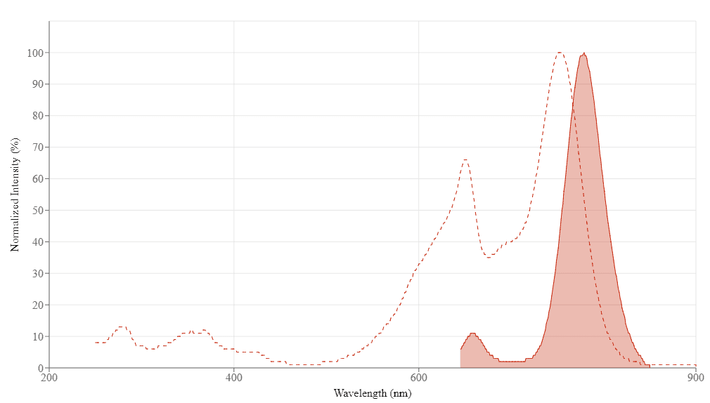
Spectral properties
| Extinction coefficient (cm -1 M -1) | 700000 |
| Excitation (nm) | 651 |
| Emission (nm) | 779 |
Product Family
| Name | Excitation (nm) | Emission (nm) | Extinction coefficient (cm -1 M -1) |
| Buccutite™ Rapid PE-Cy7 Tandem Antibody Labeling Kit *Microscale Optimized for Labeling 100 ug Antibody Per Reaction* | 565 | 778 | 1960000 |
| Buccutite™ Rapid PE-Cy7 Tandem Antibody Labeling Kit *Microscale Optimized for Labeling 25 ug Antibody Per Reaction* | 565 | 778 | 1960000 |
| Buccutite™ Rapid PE-Cy7 Tandem Antibody Labeling Kit *Production Scale Optimized for Labeling 1 mg Antibody Per Reaction* | 565 | 778 | 1960000 |
Images

References
Authors: Kudryavtsev, Igor and Zinchenko, Yulia and Starshinova, Anna and Serebriakova, Maria and Malkova, Anna and Akisheva, Tatiana and Kudlay, Dmitriy and Glushkova, Anzhela and Yablonskiy, Piotr and Shoenfeld, Yehuda
Journal: Diagnostics (Basel, Switzerland) (2023)
Authors: Jensen, Holly A and Kim, Jeong
Journal: Cytometry. Part A : the journal of the International Society for Analytical Cytology (2022)
Authors: Laufer, Natalia and Ojeda, Diego and Polo, María Laura and Martinez, Ana and Pérez, Héctor and Turk, Gabriela and Cahn, Pedro and Zwirner, Norberto Walter and Quarleri, Jorge
Journal: World journal of hepatology (2017): 1073-1080
Authors: De Biasi, Sara and Gibellini, Lara and Bianchini, Elena and Nasi, Milena and Pinti, Marcello and Salvioli, Stefano and Cossarizza, Andrea
Journal: Cytometry. Part A : the journal of the International Society for Analytical Cytology (2016): 1106-1110
Authors: Kong, Y and Xu, L-P and Liu, Y-R and Qin, Y-Z and Sun, Y-Q and Wang, Y and Jiang, H and Jiang, Q and Chen, H and Chang, Y-J and Huang, X-J
Journal: Bone marrow transplantation (2015): 348-53
Authors: Elezov, D S and Kudryavtsev, I V and Arsent'ev, N A and Basin, V V and Esaulenko, E V and Semenov, A V and Totolyan, A A
Journal: Bulletin of experimental biology and medicine (2015): 238-42
Authors: Moore, Jeffrey P and Sakkal, Samy and Bullen, Michelle L and Kemp-Harper, Barbara K and Ricardo, Sharon D and Sobey, Christopher G and Drummond, Grant R
Journal: Journal of immunological methods (2013): 33-43
Authors: Degheidy, Heba A and Venzon, David J and Farooqui, Mohammed Z H and Abbasi, Fatima and Arthur, Diane C and Wilson, Wyndham H and Wiestner, Adrian and Stetler-Stevenson, M A and Marti, Gerald E
Journal: Cytometry. Part B, Clinical cytometry (2012): 67-77
Authors: Cannizzo, Elisa and Carulli, Giovanni and Del Vecchio, Luigi and Ottaviano, Virginia and Bellio, Emanuele and Zenari, Ezio and Azzarà, Antonio and Petrini, Mario and Preffer, Frederic
Journal: American journal of clinical pathology (2012): 377-86
Authors: Tölle, Angelika and Abdallah, Ziyad and Jung, Klaus and Bäumler, Hans
Journal: Journal of fluorescence (2010): 779-86