
Products
Services
Resources
Selection Guides
About
Autophagy and Mitophagy
Autophagy and mitophagy are essential catabolic processes that maintain cellular homeostasis through the degradation and recycling of cytoplasmic components, damaged organelles, and long-lived proteins. Autophagy involves the formation of double-membrane vesicles called autophagosomes that engulf cellular cargo and fuse with lysosomes for degradation, while mitophagy specifically targets dysfunctional mitochondria for selective removal. These processes play critical roles in development, aging, and disease pathogenesis, with dysregulation implicated in neurodegenerative disorders, cancer, and metabolic diseases.
AAT Bioquest offers a comprehensive suite of fluorescent probes and assay kits for monitoring autophagy and mitophagy in live cells, enabling researchers to visualize autophagosome formation, track autophagic flux, and assess mitochondrial quality control mechanisms using fluorescence microscopy, flow cytometry, and high-throughput screening platforms.
Product Line | Application | Platform | Available Fluorescence |
|---|---|---|---|
Cell Meter™ Autophagy Assay Kits | Autophagy | Fluorescence microscope, fluorescence microplate reader | Blue, green |
DAP Dyes | Autophagy | Fluorescence microscope | Green, red |
Cell Meter™ Mitochondrial Autophagy Imaging Kit | Mitophagy | Fluorescence microscope | Red |
The Cell Meter™ Autophagy Assay Kits provide complete solutions for detecting and quantifying autophagy in live cells. These kits utilize proprietary fluorescent probes that accumulate in autophagic vacuoles and related acidic vesicular compartments, enabling real-time monitoring of autophagy-associated vesicle formation and changes in autophagic activity. The assays are compatible with various induction methods including nutrient starvation, rapamycin treatment, and other autophagy-modulating compounds. Multiple fluorescence options allow for flexible experimental design and multiplexing with other cellular markers.
Fig. 1
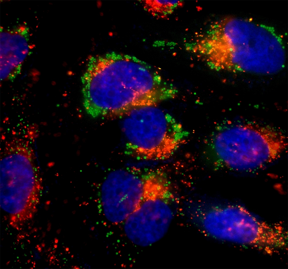
HeLa cells were incubated in 1X HBSS buffer with 5% serum to induce starvation. Following starvation, cells were treated with Autophagy Green™ (Catalog Number 23002) working solution for 20 minutes in a 37°C, 5% CO2 incubator and then washed 3 times. Nuclei were labeled with Hoechst 33342 (Catalog Number 17530). Lysosomes were labeled with LysoBrite™ Orange (Catalog Number 22657).
DAP dyes are cell-permeable fluorescent probes that selectively accumulate in autophagic vacuoles and exhibit enhanced fluorescence in lipid-rich, acidic membrane environments. These dyes enable real-time, wash-free monitoring of autophagy in live cells with minimal background fluorescence. The availability of both green and red fluorescent options allows for multiplexing with other cellular markers and co-localization studies with organelle-specific probes.
Mitochondrial autophagy (mitophagy) is a selective form of autophagy that targets damaged or dysfunctional mitochondria for lysosomal degradation. This process is critical for maintaining mitochondrial quality control and has been implicated in the pathogenesis of neurodegenerative diseases including Alzheimer's and Parkinson's disease. The Cell Meter™ Mitochondrial Autophagy Imaging Kit enables visualization of mitophagy-associated events using a red-fluorescent probe that reports mitochondrial delivery to autophagic and lysosomal compartments.
Fig. 2
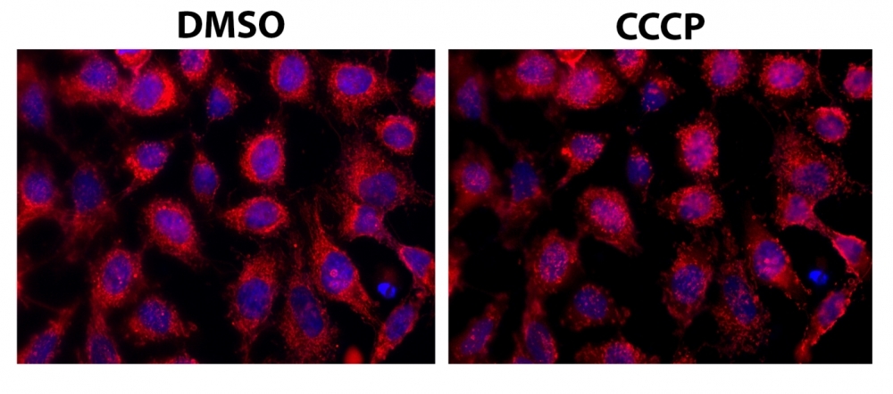
The fluorescence images of HeLa cells co-stained with Mitophagy Red™ and Hoechst 33342 (Catalog Number 17535) in a 96-well black-wall clear-bottom plate. Image was acquired before (Left) and after (Right) addition of CCCP (10 uM) for 1 minute. The cells were imaged using a fluorescence microscope with a Cy3/TRITC filter.
- Nutrient starvation (HBSS or serum-free medium)
- Rapamycin treatment (mTOR inhibition)
- Chloroquine or bafilomycin A1 (lysosomal inhibitors for flux measurement)
- CCCP or FCCP (mitochondrial uncouplers for mitophagy induction)
For comprehensive autophagy studies, consider combining autophagy probes with:
- Nuclear stains for cell identification
- Mitochondrial markers (MitoLite™ probes) for co-localization
- Lysosomal probes (LysoBrite™) for autophagosome-lysosome fusion analysis
- Viability indicators for distinguishing autophagy from cell death
This document (01.0228.251203r1) was last updated on Sat Feb 28 2026. All trademarks and registered trademarks mentioned herein are the property of their respective owners.