
Products
Services
Resources
Selection Guides
About
Mitochondria
Mitochondria are distinctive, membrane-bound organelles present within all eukaryotic cells. As the primary site of ATP production and cellular energy supply, the unique aspects of mitochondria (including separate genetic material termed mtDNA, their own protein-synthesizing machinery, and a strictly regulated membrane potential charge gradient) make them an important subject of study. Additionally, as more aspects of the aging process and other human diseases are being discovered, the role of mitochondria has been shown to be even more prominent than previously suspected.
AAT Bioquest offers a wide range of imaging technologies for investigating mitochondrial morphology and functionality in live and fixed cells. The selective labeling of live cell compartments provides a powerful method for examining cellular events in a spatial and temporal context.
| Use for | Readout | Products | |
|---|---|---|---|
| Mitochondrial Staining | Mitochondrial morphology and localization in live cells, with fixable options | Blue to near-infrared fluorescence | MitoLite™ dyes, Cell Navigator® Mitochondrion Staining Kits, and CytoFix™ Red |
| Membrane Potential (Δψₘ) | Mitochondrial health and depolarization during early apoptosis | Orange-to-green emission ratio (JC-10™) or orange intensity (TMRE and TMRM) | JC-10™, JC-1, TMRE, TMRM, and Cell Meter™ JC-10™ assay kits |
| Mitochondrial DNA | Mitochondrial DNA labeling in live cells | Blue to far-red fluorescence | MitoDNA™ dyes |
| Mitochondrial ROS | Superoxide, hydroxyl radical, hydrogen peroxide, and broad-spectrum ROS detection | Green to near-infrared fluorescence | MitoROS Brite™ and MitoROS™ indicators |
 Live HeLa cells stained with MitoLite™ Red FX600 (red), ER Green™ (green) and DAPI (blue).
MitoLite™ dyes and CytoFix™ Red label mitochondria for morphology and localization studies. They differ mainly in how well the staining is retained after fixation.
MitoLite™ dyes are a series of cell-permeable cationic dyes used to label mitochondria in live cells. At micromolar concentrations (μM), MitoLite™ dyes passively diffuse across live cell membranes and selectively accumulate in active mitochondria via the mitochondrial membrane potential (Δψₘ) gradient. Since staining is lost during cell death due to the collapse (depolarization) of the mitochondrial membrane potential, MitoLite™ dyes can also be used to monitor cell viability.
MitoLite™ dyes are available in spectral characteristics ranging from blue to near-infrared to accommodate multiplexing with other fluorescent probes. Retention after fixation varies amongst MitoLite™ dyes, with dyes such as MitoLite™ Blue FX490 being well-retained and MitoLite™ Green FM being lost after fixation. MitoLite™ dyes are key components of Cell Navigator® Mitochondrion Staining Kits. Each kit provides sufficient materials for 500 tests.
Bright, photostable fluorescence with high signal-to-noise ratios | Can be imaged without washing |
Multiple wavelength options for multiplex analysis with other fluorescent probes | Suitable for proliferating and non-proliferating cells, in suspension or adherent formats |

Fluorescence imaging of HeLa cells stained with the MitoLite™ mitochondrial dye series.
Left: HeLa cells stained with MitoLite™ Blue FX490 (Catalog Number 22674).
Center: HeLa cells stained with MitoLite™ Green FM (Catalog Number 22695), co-stained with LysoBrite™ Red (Catalog Number 22645) and Nuclear Violet™ LCS1 (Catalog Number 17543).
Right: HeLa cells stained with MitoLite™ NIR FX690 (Catalog Number 22690), labeled with iFluor® 488-Phalloidin (Catalog Number 23115) and counterstained with Nuclear Blue™ DCS1 (Catalog Number 17548).
The enhanced cellular retention of CytoFix™ Red mitochondrial stain preserves its mitochondrial localized fluorescence even after the mitochondrial membrane potential is lost during fixation. The fluorescence signal generated by this dye is well retained in mitochondria, making it convenient for long-term mitochondrial tracking. CytoFix™ Red mitochondrial stain can be used with other fluorescent conjugates and other organelle stains for multicolor analysis. It can be used for both suspension and adherent cells and readily adapted for a wide variety of fluorescence platforms.
High mitochondrial staining efficiency | Long retention after fixation for long-term mitochondrial tracking |
Labeling protocol with minimal hands-on time | Multicolor-compatible with other fluorescent conjugates and organelle stains |
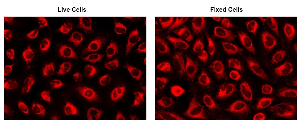 The fluorescence images of HeLa cells stained with CytoFix™ MitoRed in a 96-well black-wall clear-bottom plate. Images were acquired before (Left) and after (Right) fixation with 4% formaldehyde solution for 20 minutes at RT. The cells were imaged using a fluorescence microscope equipped with a Cy3/TRITC filter.
The mitochondrial membrane potential (Δψₘ), generated by the electron transport chain, is a key parameter necessary for healthy mitochondrial functioning. Together with the proton gradient, it generates the driving force behind mitochondrial ATP synthesis. It plays a key role in mitochondrial homeostasis through selective elimination of dysfunctional mitochondria, and is an essential component of mitochondrial calcium homeostasis.
A distinctive feature of the early stages of apoptosis is the disruption of normal mitochondrial function. A collapse in mitochondrial membrane and redox potential may induce unwanted loss of cell viability and be a cause of various pathologies. JC-10™ reports Δψₘ through the ratio of its orange and green emission, while TMRE and TMRM report it through fluorescence intensity.
Improving upon classic reagents like JC-1, JC-10™ is a potential-dependent probe used to determine Δψₘ by flow cytometry, fluorescence microscopy, and microplate-based fluorescent assays. In healthy cells, JC-10™ selectively accumulates in mitochondria generating orange J-aggregates that exhibit a broad excitation spectrum and emission maximum at 590 nm. However, in apoptotic and necrotic cells with low Δψₘ, JC-10™ diffuses out of the mitochondria and JC-10™ monomers are generated, resulting in a shift to green emission (525 nm). JC-10™ allows both qualitative and quantitative visualization of Δψₘ changes, considering the shift from orange to green fluorescence emission and the fluorescence intensity ratio, respectively.
No precipitation in aqueous buffers, for fewer artifacts and smaller assay deviations | Higher signal-to-background ratio than JC-1 |
Detects subtle Δψₘ loss better than JC-1 in all tested cell lines | Works in primary rat hepatocytes and many other cell types |
 Camptothecin induced mitochondrial membrane potential changes were measured with JC-10™ (Catalog Number 22204) and JC-1 (Catalog Number 22200) in Jurkat cells. After Jurkat cells were treated with camptothecin (10 µM) for 4 hours, JC-1 and JC-10™ dye loading solutions were added to the wells and incubated for 30 minutes. The fluorescent intensities for both J-aggregates and monomeric forms of JC-1 and JC-10™ were measured at Ex/Em = 490/525 nm and 540/590 nm with NOVOstar microplate reader (BMG Labtech).
Cell-permeable cationic rhodamines, such as TMRE and TMRM, are readily sequestered by active mitochondria, and commonly used to label mitochondria in living cells. Like JC-10™, TMRE and TMRM uptake in mitochondria is driven by the mitochondrial membrane potential. Both dyes have been used successfully for dynamic and in situ quantitative measurements, to screen for inhibitors of the mitochondrial transition pore, to assess the functionality of mitochondria in living cells, and to discriminate between viable and non-viable cell populations. These potentiometric dyes exhibit minimal self-quenching, low cytotoxicity, and reasonable photostability, and their fluorescence intensities can be measured with either a flow cytometer or fluorescence microscope. In comparison to TMRM, TMRE is slightly more hydrophobic.
Mitochondrial DNA (mtDNA) is a small circular DNA that codes for 37 genes in humans, including essential enzymes involved in oxidative phosphorylation and genes for transfer RNA (tRNA) and ribosomal RNA (rRNA). Mutations in mtDNA are linked to age-related hearing loss, diabetes, organ failure, and several types of cancer.
Common fluorescent DNA probes such as DAPI, Hoechst, or SYBR® Green lack the specificity to target mitochondria and predominantly stain cell nuclei. The MitoDNA™ dye series specifically stains mtDNA in live cells.
Cell-permeable and specific for mitochondrial DNA | Large Stokes shifts |
Excellent signal-to-noise ratio | Suitable for multiplex analysis with other fluorescent probes |
 Fluorescence response of the MitoDNA™ dye series in HeLa cells. HeLa cells stained with MitoDNA™ Blue 470 (Left, Catalog Number 22684) exhibit bright mitochondrial fluorescence when imaged by fluorescence microscopy. The MitoDNA™ dye set also includes MitoDNA™ Green 530 (Center-Left, Catalog Number 22685), MitoDNA™ Red 680 (Center-Right,Catalog Number 22688), and MitoDNA™ Red 710 (Right, Catalog Number 22689), providing a range of spectral options for multicolor visualization of mitochondrial DNA and high-contrast mitochondrial DNA imaging.
Mitochondrial reactive oxygen species (mtROS or mROS), including superoxide anion, hydrogen peroxide, and hydroxyl radicals, are primarily generated at the electron transport chain within the inner mitochondrial membrane during oxidative phosphorylation. At basal and moderate levels, mtROS regulate inflammatory responses, metabolic adaptation, gene expression, cell proliferation, and differentiation, while excessive accumulation can activate apoptosis and autophagy pathways. Their roles in tumorigenesis and in viral infection have made mtROS a therapeutic target.
MitoROS Brite™ dyes detect mitochondrial ROS in live cells. The series includes MitoROS Brite™ 570 and MitoROS Brite™ 670, which target a broad spectrum of ROS, and MitoROS Brite™ NIR 780, which selectively detects hydrogen peroxide. These cell-permeable probes accumulate in mitochondria and are rapidly oxidized by their specific ROS, producing a robust fluorescent signal.
For individual species, AAT Bioquest provides the superoxide-specific dyes MitoROS™ 520 (Catalog Number 16060) and MitoROS™ 580 (Catalog Number 16052), which are key components of the Cell Meter™ Fluorimetric Mitochondrial Superoxide Activity Assay Kits. The hydroxyl radical-selective probe MitoROS™ OH580 is the key component of the Cell Meter™ Mitochondrial Hydroxyl Radical Detection Kit (Catalog Number 16055).
Strong retention of oxidized fluorophore within live cells | Specific for defined classes of mitochondrial reactive oxygen species |
High signal-to-noise ratio | Optimization for multiplex analysis with other mitochondrial probes |
 The fluorescence response of MitoROS Brite™ NIR 780 (0.5 µM) to varying concentrations of H2O2 in HeLa cells was assessed. Fluorescence intensities were monitored using a fluorescence microscope equipped with a Cy7 filter.
This document (01.0086.211015r2) was last updated on Wed Nov 11 2026. All trademarks and registered trademarks mentioned herein are the property of their respective owners.
Mitochondria