logo
AAT Bioquest

Cell Meter™ IX830 fixable viability dye

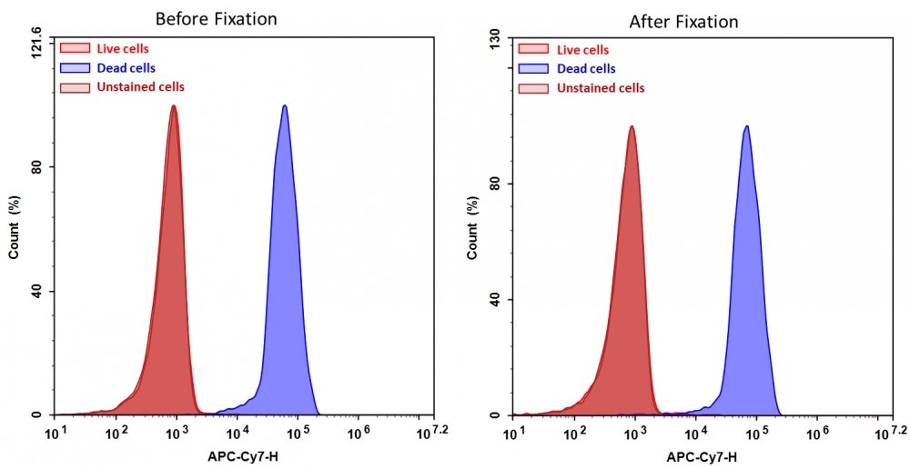
Discrimination and exclusion of dead cells from live cells allows cleaner separation and identification of cell populations. Cell Meter™ fixable viability dyes are a large family of cell-impermeable fluorescent viability dyes that are optimized to match the major excitation lasers of common flow cytometers, such as 350, 405, 488, 633 and 647 nm. These dyes are impermeant to live cells but permeant to cells with compromised membranes. They irreversibly react with amine- and thiol-containing proteins and other cellular components. Since dead or fixed cells with a compromised membrane more readily react with Cell Meter™ fixable cell stains, thus stain brighter than live cells with an intact membrane, these dyes can be used to assess live vs. dead status of mammalian cells. There are a few factors to be considered when using these dyes, e.g., the titration of each dye to ensure that live cells have minimal to no staining. Cell Meter™ IX830 fixable cell stain is optimized to be excited with the IR laser at 808 nm with emission at 830 nm. Compared to other commercially similar viability dyes, this fixable viability dye is much more robust and stable.
Example protocol

AT A GLANCE

Protocol summary
  1. Prepare samples in HHBS buffer (0.5 mL/assay).

  2. Add Cell Meter™ IX830 to the cell suspension.

  3. Stain the cells at room temperature for 20 - 60 minutes.

  4. Wash the cells.

  5. Fix the cells (optional).

  6. Examine the sample with flow cytometer using 780/50 nm filter (APC-Cy7 channel) and/or IR filters.

Important

Thaw at room temperature before starting the experiment.

PREPARATION OF STOCK SOLUTIONS

Unless otherwise noted, all unused stock solutions should be divided into single-use aliquots and stored at -20 °C after preparation. Avoid repeated freeze-thaw cycles

Cell Meter™ IX830 stock solution (500X)
  1. Add 200 µL of DMSO to the Cell Meter™ IX830 vial to make a 500X stock solution.

    Note: Any unused Cell Meter™ IX830 stock solution should be divided into single-use aliquots and stored at -20°C. Avoid repeated freeze/thaw cycles.

SAMPLE EXPERIMENTAL PROTOCOL

  1. Treat cells as desired.

  2. Wash cells once with HHBS or the azide- and serum/protein-free buffer of your choice.

  3. Resuspend cells at 5 - 10 × 10 6/mL in HHBS or in the azide- and serum/protein-free buffer of your choice.

  4. Add 1 µL of 500X Cell Meter™ IX830 stock solution to 0.5 mL of cells/assay and mix it well.

  5. Incubate at room temperature or 37°, 5% CO2 incubator for 20 - 60 minutes, protected from light.

    Note: The optimal stain concentrations and incubation time should be experimentally determined for different cell lines.

  6. Wash cells twice and resuspend cells with HHBS or the buffer of your choice.

  7. Fix cells as desired (optional).

  8. Analyze cells with a flow cytometer using 780/50 nm filter (APC-Cy7 channel) or IR filters.

Spectrum

References

View all 50 references: Citation Explorer
Hsa_circ_0011385 accelerates the progression of thyroid cancer by targeting miR-361-3p.
Authors: Xia, Fada and Chen, Yong and Jiang, Bo and Bai, Ning and Li, Xinying
Journal: Cancer cell international (2020): 49
Immunomodulatory effects of some Namibian plants traditionally used for treating inflammatory diseases.
Authors: Du Preez, C I and Gründemann, C and Mumbengegwi, D R and Huber, R
Journal: Journal of ethnopharmacology (2020): 112683
UHPLC-Q/Orbitrap/MS/MS fingerprinting and antitumoral effects of Prosopis strombulifera (LAM.) BENTH. queous extract on allograft colorectal and melanoma cancer models.
Authors: Persia, Fabio Andrés and Troncoso, Mariana Elizabeth and Rinaldini, Estefanía and Simirgiotis, Mario and Tapia, Alejandro and Bórquez, Jorge and Mackern-Oberti, Juan Pablo and Hapon, María Belén and Gamarra-Luques, Carlos
Journal: Heliyon (2020): e03353
Lignins isolated from Prickly pear cladodes of the species Opuntia fícus-indica (Linnaeus) Miller and Opuntia cochenillifera (Linnaeus) Miller induces mice splenocytes activation, proliferation and cytokines production.
Authors: da Cruz Filho, Iranildo José and da Silva Barros, Bárbara Rafaela and de Souza Aguiar, Lethícia Maria and Navarro, Claudia Daniele Carvalho and Ruas, Juliana Silveira and de Lorena, Virgínia Maria Barros and de Moraes Rocha, George Jackson and Vercesi, Aníbal Eugênio and de Melo, Cristiane Moutinho Lagos and Maior, Ana Maria Souto
Journal: International journal of biological macromolecules (2019): 1331-1339
Research on the effect and mechanism of antimicrobial peptides HPRP-A1/A2 work against Toxoplasma gondii infection.
Authors: Liu, Ran and Ni, Yangyue and Song, Jingwei and Xu, Zhipeng and Qiu, Jingfan and Wang, Lijuan and Zhu, Yuxiao and Huang, Yibing and Ji, Minjun and Chen, Yuxin
Journal: Parasite immunology (2019): e12619
Page updated on December 11, 2024

Ordering information

Price
Unit size
Catalog Number22529
Quantity
Add to cart

Additional ordering information

Telephone1-800-990-8053
Fax1-800-609-2943
Emailsales@aatbio.com
InternationalSee distributors
Bulk requestInquire
Custom sizeInquire
Technical SupportContact us
Purchase orderSend to sales@aatbio.com
ShippingStandard overnight for United States, inquire for international
Request quotation

Physical properties

Solvent

DMSO

Spectral properties

Excitation (nm)

811

Emission (nm)

822

Storage, safety and handling

H-phraseH303, H313, H333
Hazard symbolXN
Intended useResearch Use Only (RUO)
R-phraseR20, R21, R22

Storage

Freeze (< -15 °C); Minimize light exposure
UNSPSC12171501

Platform

Flow cytometer

Excitation634 laser or IR lasers
Emission780, 50 nm or IR filters
Instrument specification(s)APC, Cy7 or IR filters
Detection of Jurkat cell viability by Cell Meter&trade; fixable viability dye. Jurkat cells were treated and stained with&nbsp;Cell Meter&trade; IX830 (Cat#22529), and then fixed in 3.7% formaldehyde and analyzed by flow cytometry. &nbsp;The dead cell population (Blue peak)&nbsp; is easily distinguished from the live cell population (Red peak)&nbsp; with APC-Cy7 channel, and nearly identical results were obtained before and after fixation.