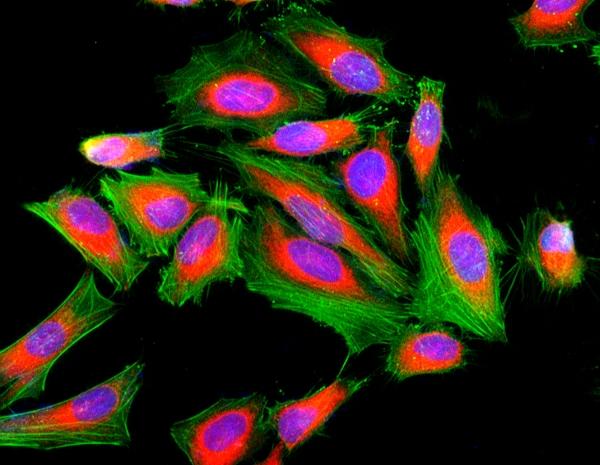
Cell Navigator® F-Actin Labeling Kit *Green Fluorescence*
Our Cell Navigator® fluorescence imaging kits are a set of fluorescence imaging tools for labeling sub-cellular organelles such as membranes, lysosomes, mitochondria and nuclei etc. The selective labeling of live cell compartments provides a powerful method for studying cellular events in a spatial and temporal context. This particular kit is designed to label F-actins of fixed cells in green fluorescence. The kit uses a green fluorescent phalloidin conjugate that is selectively bound to F-actins. This green fluorescent phalloidin conjugate is a high-affinity probe for F-actins with much higher photostability than the fluorescein-phalloidin conjugates. Used at nanomolar concentrations, phallotoxins are convenient probes for labeling, identifying and quantitating F-actins in formaldehyde-fixed and permeabilized tissue sections, cell cultures or cell-free experiments. The labeling protocol is robust, requiring minimal hands-on time. The kit provides all the essential components with an optimized staining protocol.
Example protocol
AT A GLANCE
Protocol summary
- Prepare samples (microplate wells)
- Remove the liquid from the plate
- Add 100 µL/well of iFluor™ 488-Phalloidin working solution
- Stain the cells at RT for 15 to 60 minutes
- Wash the cells
- Examine the specimen under fluorescence microscope at Ex/Em = 490/520 nm (FITC filter set)
Important notes
Thaw all the components at room temperature before starting the experiment.
PREPARATION OF WORKING SOLUTION
Add 10 μL of iFluor™ 488-Phalloidin (Component A) to 10 mL of Labeling Buffer (Component B) to make 1X iFluor™ 488-Phalloidin working solution. Protect from light. Note: Different cell types might be stained differently. The concentration of iFluor™ 488-Phalloidin working solution should be prepared accordingly.
For guidelines on cell sample preparation, please visit
https://www.aatbio.com/resources/guides/cell-sample-preparation.html
SAMPLE EXPERIMENTAL PROTOCOL
- Perform formaldehyde fixation. Incubate the cells with 3.0% – 4.0% formaldehyde in PBS at room temperature for 10 – 30 minutes. Note: Avoid any methanol containing fixatives since methanol can disrupt actin during the fixation process. The preferred fixative is methanol-free formaldehyde.
- Rinse the fixed cells 2 – 3 times in PBS.
- Optional: Add 0.1% Triton X-100 in PBS into fixed cells for 3 to 5 minutes to increase permeability. Rinse the cells 2 – 3 times in PBS.
- Add 100 µL/well (96-well plate) of iFluor™ 488-Phalloidin working solution into the fixed cells.
- Stain the cells at room temperature for 15 to 60 minutes.
- Rinse cells gently with PBS 2 to 3 times to remove excess dye before plate sealing.
- Image cells using a fluorescence microscope with FITC filter set (Ex/Em = 490/520 nm).
Spectrum
Product family
| Name | Excitation (nm) | Emission (nm) | Extinction coefficient (cm -1 M -1) | Quantum yield | Correction Factor (260 nm) | Correction Factor (280 nm) |
| Cell Navigator® F-Actin Labeling Kit *Blue Fluorescence* | 345 | 450 | 200001 | 0.951 | 0.83 | 0.23 |
| Cell Navigator® F-Actin Labeling Kit *Orange Fluorescence* | 541 | 557 | 1000001 | 0.671 | 0.25 | 0.15 |
| Cell Navigator® F-Actin Labeling Kit *Red Fluorescence* | 587 | 603 | 2000001 | 0.531 | 0.05 | 0.04 |
Citations
View all 47 citations: Citation Explorer
TNF-$\alpha$ induces premature senescence in tendon stem cells via the NF-$\kappa$B and p53/p21/cyclin E/CDK2 signaling pathways
Authors: Guo, Hua and Cao, Haixia and Lu, Qian and Gu, Zhifeng and Feng, Guijuan
Journal: International Journal of Molecular Medicine (2025): 1--14
Authors: Guo, Hua and Cao, Haixia and Lu, Qian and Gu, Zhifeng and Feng, Guijuan
Journal: International Journal of Molecular Medicine (2025): 1--14
NINJ1 regulates plasma membrane fragility under mechanical strain
Authors: Zhu, Yunfeng and Xiao, Fang and Wang, Yiling and Wang, Yufang and Li, Jialin and Zhong, Dongmei and Huang, Zhilei and Yu, Miao and Wang, Zhirong and Barbara, Joshua and others,
Journal: Nature (2025): 1--3
Authors: Zhu, Yunfeng and Xiao, Fang and Wang, Yiling and Wang, Yufang and Li, Jialin and Zhong, Dongmei and Huang, Zhilei and Yu, Miao and Wang, Zhirong and Barbara, Joshua and others,
Journal: Nature (2025): 1--3
Engineered Extracellular Vesicles Derived from Juvenile Mice Enhance Mitochondrial Function in the Aging Bone Microenvironment and Achieve Rejuvenation
Authors: Zheng, Jiaqian and Ren, Yipeng and Ke, Junhua and Zhu, Guanglin and Wang, Zhen and Shi, Xuetao and Wang, Yingjun
Journal: ACS nano (2025)
Authors: Zheng, Jiaqian and Ren, Yipeng and Ke, Junhua and Zhu, Guanglin and Wang, Zhen and Shi, Xuetao and Wang, Yingjun
Journal: ACS nano (2025)
Biphasic Calcium Phosphate Ceramic Scaffold Composed of Zinc Doped $\beta$-Tricalcium Phosphate and Silicon Doped Hydroxyapatite for Bone Tissue Engineering
Authors: Fan, Jiajia and Yuan, Xinyuan and Lu, Teliang and Ye, Jiandong
Journal: ACS Applied Bio Materials (2024)
Authors: Fan, Jiajia and Yuan, Xinyuan and Lu, Teliang and Ye, Jiandong
Journal: ACS Applied Bio Materials (2024)
Evolution of phase, morphology, physicochemical properties, and biological properties of HA ceramic with the increase of crystallinity
Authors: Zhang, Luhui and Liang, Xinzhi and Chen, Ji and Kang, Zhengyang and Ye, Jiandong and Xie, Denghui
Journal: Ceramics International (2024)
Authors: Zhang, Luhui and Liang, Xinzhi and Chen, Ji and Kang, Zhengyang and Ye, Jiandong and Xie, Denghui
Journal: Ceramics International (2024)
References
View all 42 references: Citation Explorer
Velocity distributions of single F-actin trajectories from a fluorescence image series using trajectory reconstruction and optical flow mapping
Authors: von Wegner F, Ober T, Weber C, Schurmann S, Winter R, Friedrich O, Fink RH, Vogel M.
Journal: J Biomed Opt (2008): 54018
Authors: von Wegner F, Ober T, Weber C, Schurmann S, Winter R, Friedrich O, Fink RH, Vogel M.
Journal: J Biomed Opt (2008): 54018
Visualization of F-actin and G-actin equilibrium using fluorescence resonance energy transfer (FRET) in cultured cells and neurons in slices
Authors: Okamoto K, Hayashi Y.
Journal: Nat Protoc (2006): 911
Authors: Okamoto K, Hayashi Y.
Journal: Nat Protoc (2006): 911
The effect of F-actin on the relay helix position of myosin II, as revealed by tryptophan fluorescence, and its implications for mechanochemical coupling
Authors: Conibear PB, Malnasi-Csizmadia A, Bagshaw CR.
Journal: Biochemistry (2004): 15404
Authors: Conibear PB, Malnasi-Csizmadia A, Bagshaw CR.
Journal: Biochemistry (2004): 15404
Analysis of models of F-actin using fluorescence resonance energy transfer spectroscopy
Authors: Moens PD, dos Remedios CG.
Journal: Results Probl Cell Differ (2001): 59
Authors: Moens PD, dos Remedios CG.
Journal: Results Probl Cell Differ (2001): 59
Fluorescence studies of the carboxyl-terminal domain of smooth muscle calponin effects of F-actin and salts
Authors: Bartegi A, Roustan C, Kassab R, Fattoum A.
Journal: Eur J Biochem (1999): 335
Authors: Bartegi A, Roustan C, Kassab R, Fattoum A.
Journal: Eur J Biochem (1999): 335
Page updated on September 16, 2025


