logo
AAT Bioquest

iFluor® 488 PSA™ Imaging Kit with Goat Anti-Human IgG

Product key features

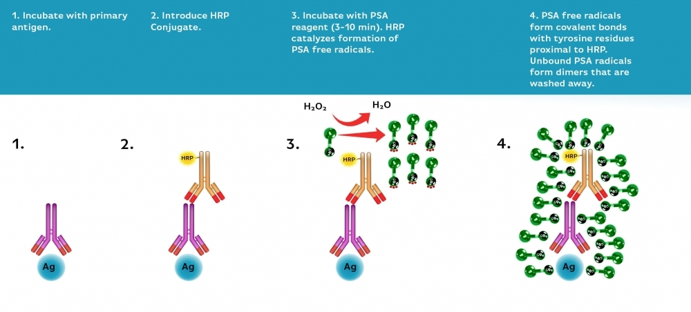
The iFluor® 488 PSA™ Imaging Kit with Goat Anti-Human IgG is designed for ultrasensitive detection of low-abundance targets in immunofluorescence assays based on Power Styramide™ Signal Amplification technology.

  • Superior sensitivity: PSA technology delivers 10–50× stronger signals than TSA reagents and >100× higher sensitivity than standard ICC/IF/IHC methods
  • Enhanced performance: iFluor® 488 dye ensures bright fluorescence, strong photostability, and excellent solubility
  • Applications: Ideal for detecting low-abundance human IgG targets in immunofluorescence and histochemistry assays
  • High performance alternative: Superior alternative to Alexa Fluor® 488 tyramide-based kits and other TSA detection systems

Product description

The iFluor® 488 PSA™ Imaging Kit with Goat Anti-Human IgG is a high-sensitivity amplification system designed for immunofluorescence and histochemistry. It utilizes Power Styramide™ Signal Amplification (PSA™), a technology that produces fluorescence signals up to 50 times stronger than conventional tyramide (TSA) reagents. Combined with the superior brightness, photostability, and water solubility of the iFluor® 488 dye, this kit enables precise and reliable detection of low-abundance targets.

This kit employs horseradish peroxidase (HRP)-mediated catalysis to covalently deposit iFluor® 488–labeled Styramide™ molecules, ensuring strong signal amplification. With optimized Goat Anti-Human IgG included, it provides specificity for detecting human IgG in cells and tissues. It is particularly useful for studies requiring sensitive immunodetection, reduced primary antibody consumption, and high reproducibility in fluorescence imaging assays.

Example protocol

AT A GLANCE

Protocol Summary
  1. Fix/permeabilize/block cells or tissue.

  2. Add primary antibody in blocking buffer.

  3. Add HRP-conjugated secondary antibody.

  4. Prepare Styramide™ working solution and apply in cells or tissue for 5-10 minutes at room temperature.

PREPARATION OF STOCK SOLUTIONS

Unless otherwise noted, all unused stock solutions should be divided into single-use aliquots and stored at -20 °C after preparation. Avoid repeated freeze-thaw cycles

Styramide™ stock solution (100X)
  1. Add 100 µL of DMSO into the vial of iFluor® 488 labeled Styramide™ conjugate (Component A) to make 100X Styramide™ stock solution.

    Note: Make single-use aliquots and store unused 100X stock solution at -20 °C in a dark place. Avoid repeat freeze-thaw cycles.

Hydrogen peroxide solution (100X)
  1. Add 10 µL of 3% hydrogen peroxide (Component E) to 90 µL of ddH2O.

    Note: Prepare the 100X H2O2 solution fresh on the day of use.

PREPARATION OF WORKING SOLUTION

Styramide working solution (1X)
  1. Every 1 mL of Reaction Buffer requires 10 µL of Styramide™ stock solution and 10 µL of H2O2 stock solution.

    Note: The Styramide™ provided is enough for 100 tests based on 100 µL of Styramide™ working solution needed per coverslip or per well in a 96-well microplate.

    Note: The Styramide™ working solution must be used within 2 hours after preparation and avoid direct exposure to light.

Secondary antibody-HRP working solution
  1. Dilute the 100X secondary antibody-HRP stock solution 1:100 in PBS with 1% BSA.

    Note: The secondary antibody-HRP provided in this kit is sufficient for 100 tests based on 100 µL HRP working solution per coverslip or per well in a 96-well microplate.

SAMPLE EXPERIMENTAL PROTOCOL

This protocol is applicable for both cells and tissues staining.

Cell fixation and permeabilization
  1. Fix the cells or tissue with 3.7% formaldehyde or paraformaldehyde, in PBS at room temperature for 20 minutes.
  2. Rinse the cells or tissue with PBS twice.
  3. Permeabilize the cells with 0.1% Triton X-100 solution for 1-5 minutes at room temperature.
  4. Rinse the cells or tissue with PBS twice.
Tissue fixation, deparaffinization and rehydration

Deparaffinize and dehydrate the tissue according to the standard IHC protocols. Perform antigen retrieval with the preferred specific solution/protocol as needed. A protocol can be found at:

https://www.aatbio.com/resources/guides/paraffin-embedded-tissue-immunohistochemistry-protocol.html  

Peroxidase labeling
  1. Optional: Quench endogenous peroxidase activity by incubating cell or tissue sample in peroxidase quenching solution (such as 3% hydrogen peroxide) for 10 minutes. Rinse with PBS twice at room temperature.
  2. Optional: If using HRP-conjugated streptavidin, it is advisable to block endogenous biotins by biotin blocking buffer (Cat. # 20047).
  3. Block with preferred blocking solution (such as PBS with 1% BSA) for 30 minutes at 4 °C.
  4. Remove blocking solution and add primary antibody diluted in recommended antibody diluent for 60 minutes at room temperature or overnight at 4 °C.
  5. Wash with PBS three times for 5 minutes each.
  6. Apply 100 µL of secondary antibody-HRP working solution to each sample and incubate for 60 minutes at room temperature.

    Note: Incubation time and concentration can be varied depending on the signal intensity.

  7. Wash with PBS three times for 5 minutes each.
Styramide labeling
  1. Prepare and apply 100 µL of Styramide™ working solution to each sample and incubate for 5-10 minutes at room temperature.

    Note: If you observe a non-specific signal, you can shorten the incubation time with Styramide. You should optimize the incubation period using positive and negative control samples at various incubation time points. Or you can use a lower concentration of Styramide in the working solution.

  2. Rinse with PBS three times.
Multiplex with primary antibodies from different species

Important: When performing multiplexing experiments with PSA™ Imaging Kits, it is essential to maintain a specific imaging sequence. To ensure clear and accurate results, it is recommended to image the targets from least to most abundant, with the nucleus counterstained last. Following this optimized imaging sequence will enhance the precision and quality of your experiment.

  1. Following Step 2 in the Styramide labeling section, you can multiplex cells or tissue samples either by employing another iFluor® PSA™ Imaging Kit or by utilizing conventional IHC/ICC protocols.

  2. For multiplexing, employ a primary antibody from a host different from the one utilized in Step 4 of the Peroxidase labeling section, along with a fluorescent label that is spectrally compatible with the initial fluorescent label.

Multiplex with primary antibodies from the same species in IHC
  1. Dilute 10X Citrate Buffer (pH 6.0) (Cat. No. 10000) 1:10 in distilled water.

  2. Following Step 2 of the Styramide labeling process, immerse the tissue in a diluted citrate buffer (pH 6.0) and heat in a microwave oven at 100% power until boiling (1–2.5 minutes).

  3. Reduce the power to 20% and microwave for an additional 15 minutes.

  4. Allow the tissue sample to cool to room temperature while it remains in the citrate buffer.

  5. Wash the sample twice with 1X PBS. Then, repeat the Peroxidase labeling and Styramide labeling steps using a primary antibody of the same species, if desired.

  6. Use an iFluor® PSA™ Imaging Kit that is spectrally compatible with the iFluor® 488 PSA™ Imaging Kit used in the first round.

Counterstain and fluorescence imaging
  1. Counterstain the cell or tissue samples as needed. AAT provides a series of nucleus counterstain reagents as listed in Table 1. Follow the instructions provided with the reagents.

  2. Mount the coverslip using a mounting medium with anti-fading properties.

    Note: To ensure optimal results, it is recommended to use either ReadiUse™ microscope mounting solution (Cat. 20009) or FluoroQuest™ TSA/PSA Antifade Mounting Medium *Optimized for Tyramide and Styramide Imaging* (Cat. 44890) instead of Vectashield® mounting media. There are instances where Vectashield® mounting media may not be suitable for certain TSA/PSA conjugates.

  3. Use the appropriate filter set to visualize the signal from the Styramide labeling.

Table 1. Recommended nucleus counterstains.

Cat#
Product Name
Ex/Em (nm)
17548
Nuclear Blue™ DCS1
350/461
17550
Nuclear Green™ DCS1
503/526
17551
Nuclear Orange™ DCS1
528/576
17552
Nuclear Red™ DCS1
642/660

Spectrum

Product family

NameExcitation (nm)Emission (nm)Extinction coefficient (cm -1 M -1)Quantum yieldCorrection Factor (260 nm)Correction Factor (280 nm)
iFluor® 350 PSA™ Imaging Kit with Goat Anti-Human IgG3454502000010.9510.830.23
iFluor® 488 PSA™ Imaging Kit with Goat Anti-Mouse IgG4915167500010.910.210.11
iFluor® 488 PSA™ Imaging Kit with Goat Anti-Rabbit IgG4915167500010.910.210.11
iFluor® 555 PSA™ Imaging Kit with Goat Anti-Human IgG55757010000010.6410.230.14
iFluor® 570 PSA™ Imaging Kit with Goat Anti-Human IgG55757012000010.7110.0490.074
iFluor® 594 PSA™ Imaging Kit with Goat Anti-Human IgG58760320000010.5310.050.04
iFluor® 647 PSA™ Imaging Kit with Goat Anti-Human IgG65667025000010.2510.030.03

References

View all 50 references: Citation Explorer
A protocol for multiplex immunofluorescence staining with a fluorescent tyramide signal amplification system, FT-GO.
Authors: Yamauchi, Kenta and Koike, Masato and Hioki, Hiroyuki
Journal: Anatomical science international (2025)
Aptamer-Based Activatable Tyramide Signal Amplification for Low-Background Detection of SARS-CoV-2 Nucleocapsid Protein.
Authors: Huang, Zhiyong and Du, Ziyan and Li, Juan and Han, Da and He, Jiaxuan and Yang, Yunben and Wang, Dan and Liang, Yu and Yang, Yunshan and Peng, Ruizi and Tan, Weihong
Journal: Analytical chemistry (2025): 328-336
A three dimensional immunolabeling method with peroxidase-fused nanobodies and fluorochromized tyramide-glucose oxidase signal amplification.
Authors: Yamauchi, Kenta and Koike, Masato and Hioki, Hiroyuki
Journal: Communications biology (2025): 903
The potential of ALFA-tag and tyramide-based fluorescence signal amplification to expand the CRISPR-based DNA imaging toolkit.
Authors: Potlapalli, Bhanu Prakash and Fuchs, Jörg and Rutten, Twan and Meister, Armin and Houben, Andreas
Journal: Journal of experimental botany (2024): 6244-6257
Protein and lipid expansion microscopy with trypsin and tyramide signal amplification for 3D imaging.
Authors: Wang, Ueh-Ting Tim and Tian, Xuejiao and Liou, Yae-Huei and Lee, Sue-Ping and Hu, Hsiao-Tang and Lu, Chieh-Han and Lin, Po-Ting and Cheng, Ya-Jen and Chen, Peilin and Chen, Bi-Chang
Journal: Scientific reports (2023): 21922
Page updated on September 13, 2025

Ordering information

Price
Unit size
Catalog Number45183
Quantity
Add to cart

Additional ordering information

Telephone1-800-990-8053
Fax1-800-609-2943
Emailsales@aatbio.com
InternationalSee distributors
Bulk requestInquire
Custom sizeInquire
Technical SupportContact us
Purchase orderSend to sales@aatbio.com
ShippingStandard overnight for United States, inquire for international
Request quotation

Spectral properties

Correction Factor (260 nm)

0.21

Correction Factor (280 nm)

0.11

Extinction coefficient (cm -1 M -1)

750001

Excitation (nm)

491

Emission (nm)

516

Quantum yield

0.91

Storage, safety and handling

H-phraseH303, H313, H333
Hazard symbolXN
Intended useResearch Use Only (RUO)
R-phraseR20, R21, R22

Platform

Fluorescence microscope

ExcitationFITC filter set
EmissionFITC filter set
Recommended plateBlack wall, clear bottom

Components

Power Styramide™ Signal Amplification (PSA™) system enables ultrasensitive detection of low-abundance targets in cells and tissues. PSA utilizes horseradish peroxidase (HRP)-mediated catalysis to deposit iFluor® dye–labeled Styramide™ molecules covalently, producing up to 10–50× stronger signals than conventional tyramide (TSA) reagents and >100× higher sensitivity than standard ICC/IF/IHC. The increased reactivity of PSA radicals, combined with the brightness and stability of iFluor® dyes, ensures faster, stronger, and more reliable fluorescence amplification compared to TSA.