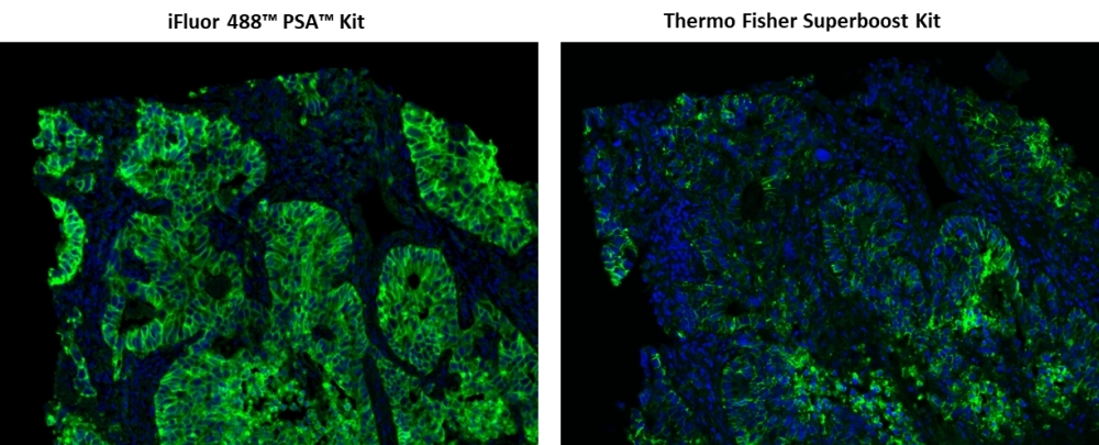
iFluor® 488 PSA™ Imaging Kit with Goat Anti-Rabbit IgG
Ordering information
Price | |
Catalog Number | |
Unit Size | |
Quantity |
Additional ordering information
Telephone | 1-800-990-8053 |
Fax | 1-800-609-2943 |
sales@aatbio.com | |
Quotation | Request |
International | See distributors |
Shipping | Standard overnight for United States, inquire for international |
Spectral properties
Correction Factor (260 nm) | 0.21 |
Correction Factor (280 nm) | 0.11 |
Extinction coefficient (cm -1 M -1) | 750001 |
Excitation (nm) | 491 |
Emission (nm) | 516 |
Quantum yield | 0.91 |
Storage, safety and handling
H-phrase | H303, H313, H333 |
Hazard symbol | XN |
Intended use | Research Use Only (RUO) |
R-phrase | R20, R21, R22 |
UNSPSC | 12352200 |
Overview | ![]() ![]() |
See also: Antibodies and Proteomics, Antibody and Protein Labeling, Bioconjugation, Cell Structures and Organelles, Nucleus, DNA and RNA Quantitation, Fluorescent in situ hybridization (FISH), Horseradish Peroxidase (HRP) and Poly-HRP, Immunohistochemistry (IHC), Physiological Probes, Power Styramide™ Signal Amplification (PSA™)
Correction Factor (260 nm) 0.21 | Correction Factor (280 nm) 0.11 | Extinction coefficient (cm -1 M -1) 750001 | Excitation (nm) 491 | Emission (nm) 516 | Quantum yield 0.91 |
Power Styramide™ Signal Amplification (PSA™) system is one of the most sensitive methods that can detect extremely low-abundance targets in cells and tissues with improved fluorescence signal 10-50 times higher than the widely used tyramide (TSA) reagents. In combination with our superior iFluor® dyes that have higher florescence intensity, increased photostability and enhanced water solubility, the iFluor® dye-labeled Styramide™ conjugates can generate fluorescence signal with significantly higher precision and sensitivity (more than 100 times) than standard ICC/IF/IHC. PSA utilizes the catalytic activity of horseradish peroxidase (HRP) for covalent deposition of fluorophores in situ. PSA radicals have much higher reactivity than tyramide radicals, making the PSA system much faster, more robust and sensitive than the traditional TSA reagents. Compared to tyramide reagents, the Styramide™ conjugates have ability to label the target at higher efficiency and thus generate significantly higher fluorescence signal. Styramide™ conjugates also allow significantly less consumption of primary antibody compared to standard directly conjugate method or tyramide amplification with the same level of sensitivity. iFluor® 488 PSA kit is a much superior replacement for Alexa Fluor 488 tyramide-based kit or other spectrally similar fluorescent tyramide or TSA kits.
Platform
Fluorescence microscope
Excitation | FITC filter set |
Emission | FITC filter set |
Recommended plate | Black wall/clear bottom |
Instrument specification(s) | FITC filter set |
Components
Example protocol
AT A GLANCE
Protocol Summary
- Fix/permeabilize/block cells or tissue
- Add primary antibody in blocking buffer
- Add HRP-conjugated secondary antibody
- Prepare Styramide™ working solution and apply in cells or tissue for 5-10 minutes at room temperature
PREPARATION OF STOCK SOLUTIONS
Unless otherwise noted, all unused stock solutions should be divided into single-use aliquots and stored at -20 °C after preparation. Avoid repeated freeze-thaw cycles.
Note Make single use aliquots, and store unused 100X stock solution at 2-8 oC in dark place and avoid repeat freeze-thaw cycles.
Note Prepare the 100X H2O2 solution fresh on the day of use.
1. Styramide™ stock solution (100X)
Add 100 µL of DMSO into the vial of iFluor™ 488-labeled Styramide™ conjugate (Component A) to make 100X Styramide™ stock solution.Note Make single use aliquots, and store unused 100X stock solution at 2-8 oC in dark place and avoid repeat freeze-thaw cycles.
2. H2O2 solution (100X)
Add 1 mL of 3% hydrogen peroxide (Component E) to 9 mL of ddH2O.Note Prepare the 100X H2O2 solution fresh on the day of use.
PREPARATION OF WORKING SOLUTION
1. Styramide working solution (1X)
Every 1 mL of Reaction Buffer requires 10 µL of Styramide™ stock solution and 10 µL of H2O2 stock solution.Note The Styramide™ provided is enough for 100 tests based on 100 µL of Styramide™ working solution needed per coverslip or per well in a 96-well microplate.
Note The Styramide™ working solution must be used within 2 hours after preparation and avoid direct exposure to light.
2. Secondary antibody-HRP working solution
Dilute the 100X secondary antibody-HRP stock solution 1:100 in PBS with 1% BSA.Note The secondary antibody-HRP provided in this kit is sufficient for 100 tests based on 100 µL HRP working solution per coverslip or per well in a 96-well microplate.
SAMPLE EXPERIMENTAL PROTOCOL
This protocol is applicable for both cells and tissues staining.
Cell fixation and permeabilization
- Fix the cells or tissue with 3.7% formaldehyde or paraformaldehyde, in PBS at room temperature for 20 minutes.
- Rinse the cells or tissue with PBS twice.
- Permeabilize the cells with 0.1% Triton X-100 solution for 1-5 minutes at room temperature.
- Rinse the cells or tissue with PBS twice.
Tissue fixation, deparaffinization and rehydration
Deparaffinize and dehydrate the tissue according to the standard IHC protocols. Perform antigen retrieval with preferred specific solution/protocol as needed.Protocol can be found at https://www.aatbio.com/resources/guides/paraffin-embedded-tissueimmunohistochemistry-protocol.html
Peroxidase labeling
- Optional: Quench endogenous peroxidase activity by incubating cell or tissue sample in peroxidase quenching solution (such as 3% hydrogen peroxide) for 10 minutes. Rinse with PBS twice at room temperature.
- Optional: If using HRP-conjugated streptavidin, it is advisable to block endogenous biotins by biotin blocking buffer.
- Block with preferred blocking solution (such as PBS with 1% BSA) for 30 minutes at 4 °C.
- Remove blocking solution and add primary antibody diluted in recommended antibody diluent for 60 minutes at room temperature or overnight at 4 °C.
- Wash with PBS three times for 5 minutes each.
- Apply 100 µL of secondary antibody-HRP working solution to each sample and incubate for 60 minutes at room temperature.
Note Incubation time and concentration can be varied depending on the signal intensity. - Wash with PBS three times for 5 minutes each.
Styramide labeling
- Prepare and apply 100 µL of Styramide™ working solution to each sample and incubate for 5-10 minutes at room temperature.
Note If you observe non-specific signal, you can shorten the incubation time with Styramide. You should optimize the incubation period using positive and negative control samples at various incubation time points. Or you can use lower concentration of Styramide in the working solution. - Rinse with PBS three times.
Counterstain and fluorescence imaging
- Counterstain the cell or tissue samples as needed. AAT provides a series of nucleus counterstain reagents as listed in Table 1. Follow the instruction provided with the reagents.
- Mount the coverslip using a mounting medium with anti-fading properties.
- Use the appropriate filter set to visualize the signal from the Styramide labeling.
Cat# | Product Name | Ex/Em (nm) |
17548 | Nuclear Blue™ DCS1 | 350/461 |
17550 | Nuclear Green™ DCS1 | 503/526 |
17551 | Nuclear Orange™ DCS1 | 528/576 |
17552 | Nuclear Red™ DCS1 | 642/660 |
Spectrum
Open in Advanced Spectrum Viewer


Spectral properties
Correction Factor (260 nm) | 0.21 |
Correction Factor (280 nm) | 0.11 |
Extinction coefficient (cm -1 M -1) | 750001 |
Excitation (nm) | 491 |
Emission (nm) | 516 |
Quantum yield | 0.91 |
Product Family
Name | Excitation (nm) | Emission (nm) | Extinction coefficient (cm -1 M -1) | Quantum yield | Correction Factor (260 nm) | Correction Factor (280 nm) |
iFluor® 350 PSA™ Imaging Kit with Goat Anti-Rabbit IgG | 345 | 450 | 200001 | 0.951 | 0.83 | 0.23 |
iFluor® 555 PSA™ Imaging Kit with Goat Anti-Rabbit IgG | 557 | 570 | 1000001 | 0.641 | 0.23 | 0.14 |
iFluor® 594 PSA™ Imaging Kit with Goat Anti-Rabbit IgG | 588 | 604 | 1800001 | 0.531 | 0.05 | 0.04 |
iFluor® 647 PSA™ Imaging Kit with Goat Anti-Rabbit IgG | 656 | 670 | 2500001 | 0.251 | 0.03 | 0.03 |
iFluor 488™ PSA™ Imaging Kit with Goat Anti-Mouse IgG | 491 | 516 | 750001 | 0.91 | 0.21 | 0.11 |
Images

Figure 1. Fluorescence IHC of formaldehyde-fixed, paraffin-embedded human lung adenocarcinoma using two imaging signal amplification kits. Tissue sections were stained with rabbit anti-EpCam antibody and then followed PSA™ method using iFluor® 488 PSA™ Imaging Kit with goat anti-rabbit IgG (Cat#45205) or Thermo Fisher Alexa Fluor® 488 Superboost Kit respectively. Cell nucleus were stained with Nuclear Blue™ DCS1 (Cat#17548).

Figure 2. Sequential immunostaining of formaldehyde-fixed, paraffin-embedded human lung adenocarcinoma using iFluor® PSA™ Imaging kits. EpCam were labeled with rabbit anti-EpCam antibodies and iFluor® 488 PSA™ Imaging Kit with goat anti-rabbit IgG, followed by washing. Pan-Keratin were labeled with mouse anti-pan Keratin antibodies and iFluor® 555 PSA™ Imaging Kit with goat anti-mouse IgG. Nuclei were labeled with DAPI. Images were acquired on a confocal microscope.

Figure 3. Sensitivity of Styramide™ Super Signal Amplification Kits. (A) HeLa cells were fixed, permeabilized and labeled with various concentrations of rabbit anti-Tubulin primary antibody. The manufacturer recommendation was 1:500 dilution. Cells were then stained with Goat anti-Rabbit IgG secondary antibody directly conjugated with Alexa Fluor® 488, or by amplified methods using a HRP-labeled Goat anti-Rabbit IgG secondary antibody followed by Alexa Fluor® 488 tyramide or iFluor® 488 Styramide™ (Cat#45205), respectively. Fluorescence images were taken using the FITC filter set and analyzed with the same exposure time. (B) Relative fluorescence signal intensity was measured and compared between different detection methods.

Figure 4. Power Styramide™ Signal Amplification (PSA™) system is one of the most sensitive methods that can detect extremely low-abundance targets in cells and tissues with improved fluorescence signal 10-50 times higher than the widely used tyramide (TSA) reagents. In combination with our superior iFluor® dyes that have higher florescence intensity, increased photostability and enhanced water solubility, the iFluor® dye-labeled Styramide™ conjugates can generate fluorescence signal with significantly higher precision and sensitivity (more than 100 times) than standard ICC/IF/IHC. PSA utilizes the catalytic activity of horseradish peroxidase (HRP) for covalent deposition of fluorophores in situ. PSA radicals have much higher reactivity than tyramide radicals, making the PSA system much faster, more robust and sensitive than the traditional TSA reagents.
Citations
View all 1 citations: Citation Explorer
CircCCNB1 silencing acting as a miR-106b-5p sponge inhibited GPM6A expression to promote HCC progression by enhancing DYNC1I1 expression and activating the AKT/ERK signaling pathway
Authors: Liu, Yan-ming and Cao, Yue and Zhao, Ping-sen and Wu, Liang-yin and Lu, Ya-min and Wang, Yu-long and Zhao, Jia-feng and Liu, Xin-guang
Journal: International journal of biological sciences (2022): 637
Authors: Liu, Yan-ming and Cao, Yue and Zhao, Ping-sen and Wu, Liang-yin and Lu, Ya-min and Wang, Yu-long and Zhao, Jia-feng and Liu, Xin-guang
Journal: International journal of biological sciences (2022): 637