logo
AAT Bioquest

mFluor™ UV780 SE

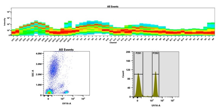
mFluor™ UV780 SE is a UV-excitable, amine-reactive fluorescent dye specifically engineered for multicolor flow cytometry applications. As part of the mFluor™ UV series, it features a large Stokes shift and is optimally excited by UV lasers at 350 nm. Its extended emission into the near-infrared region enables expanded panel design by occupying a rarely used spectral window. mFluor™ UV780 SE exhibits fluorescence excitation and emission maxima at ~350 nm and ~780 nm, respectively, making it one of the most red-shifted dyes excitable by UV light. The dye reacts efficiently with primary amines on proteins (>10 kDa), such as antibodies, providing a robust tool for custom conjugation. Its distinct spectral profile is highly compatible with spectral flow cytometers for complex immunophenotyping assays.

Example protocol

PREPARATION OF STOCK SOLUTIONS

Unless otherwise noted, all unused stock solutions should be divided into single-use aliquots and stored at -20 °C after preparation. Avoid repeated freeze-thaw cycles

Protein stock solution (Solution A)

Mix 100 µL of a reaction buffer (e.g., 1 M  sodium carbonate solution or 1 M phosphate buffer with pH ~9.0) with 900 µL of the target protein solution (e.g. antibody, protein concentration >2 mg/mL if possible) to give 1 mL protein labeling stock solution.

Note: The pH of the protein solution (Solution A) should be 8.5 ± 0.5. If the pH of the protein solution is lower than 8.0, adjust the pH to the range of 8.0-9.0 using 1 M  sodium bicarbonate solution or 1 M pH 9.0 phosphate buffer.

Note: The protein should be dissolved in 1X phosphate buffered saline (PBS), pH 7.2-7.4. If the protein is dissolved in Tris or glycine buffer, it must be dialyzed against 1X PBS, pH 7.2-7.4, to remove free amines or ammonium salts (such as ammonium sulfate and ammonium acetate) that are widely used for protein precipitation.

Note: Impure antibodies or antibodies stabilized with bovine serum albumin (BSA) or gelatin will not be labeled well. The presence of sodium azide or thimerosal might also interfere with the conjugation reaction. Sodium azide or thimerosal can be removed by dialysis or spin column for optimal labeling results.

Note: The conjugation efficiency is significantly reduced if the protein concentration is less than 2 mg/mL. For optimal labeling efficiency the final protein concentration range of 2-10 mg/mL is recommended.

mFluor™ UV780 SE stock solution (Solution B)

Add anhydrous DMSO into the vial of mFluor™ UV780 SE to make a 10 mM stock solution. Mix well by pipetting or vortex.

Note: Prepare the dye stock solution (Solution B) before starting the conjugation. Use promptly. Extended storage of the dye stock solution may reduce the dye activity. Solution B can be stored in freezer for two weeks when kept from light and moisture. Avoid freeze-thaw cycles.

SAMPLE EXPERIMENTAL PROTOCOL

This labeling protocol was developed for the conjugate of Goat anti-mouse IgG with mFluor™ UV780 SE. You might need further optimization for your particular proteins.

Note: Each protein requires distinct dye/protein ratio, which also depends on the properties of dyes. Over labeling of a protein could detrimentally affects its binding affinity while the protein conjugates of low dye/protein ratio gives reduced sensitivity.

Run conjugation reaction
  1. Use 10:1 molar ratio of Solution B (dye)/Solution A (protein) as the starting point:  Add 5 µL of the dye stock solution (Solution B), assuming the dye stock solution is 10 mM) into the vial of the protein solution (95 µL of Solution A) with effective shaking. The concentration of the protein is ~0.05 mM assuming the protein concentration is 10 mg/mL and the molecular weight of the protein is ~200KD.

    Note: We recommend to use 10:1 molar ratio of Solution B (dye)/Solution A (protein). If it is too less or too high, determine the optimal dye/protein ratio at 5:1, 15:1 and 20:1 respectively.

  2. Continue to rotate or shake the reaction mixture at room temperature for 30-60 minutes.
Purify the conjugation

The following protocol is an example of dye-protein conjugate purification by using a Sephadex G-25 column.

  1. Prepare Sephadex G-25 column according to the manufacture instruction.
  2. Load the reaction mixture (From "Run conjugation reaction") to the top of the Sephadex G-25 column.
  3. Add PBS (pH 7.2-7.4) as soon as the sample runs just below the top resin surface.
  4. Add more PBS (pH 7.2-7.4) to the desired sample to complete the column purification. Combine the fractions that contain the desired dye-protein conjugate.

    Note: For immediate use, the dye-protein conjugate need be diluted with staining buffer, and aliquoted for multiple uses.

    Note: For longer term storage, dye-protein conjugate solution need be concentrated or freeze dried.

Spectrum

Product family

NameExcitation (nm)Emission (nm)Extinction coefficient (cm -1 M -1)Quantum yieldCorrection Factor (260 nm)Correction Factor (280 nm)
mFluor™ UV375 SE3513873000010.9410.0990.138
mFluor™ UV420 SE353421750001---
mFluor™ UV455 SE3574612000010.4210.6510.406
mFluor™ UV460 SE3584561500010.8610.350.134
mFluor™ UV520 SE503524800001-0.4950.518
mFluor™ UV540 SE5425609000010.3510.6340.463
mFluor™ UV595 SE5815941400001-0.1810.168
mFluor™ UV610 SE5896099000010.250.9490.904

References

View all 7 references: Citation Explorer
Photoswitch-Based Fluorescence Encoding of Microspheres in a Limited Spectral Window for Multiplexed Detection.
Authors: Guo, Chao and Zhai, Jingying and Wang, Yifu and Du, Xinfeng and Wang, Zige and Xie, Xiaojiang
Journal: Analytical chemistry (2022): 1531-1536
Fluorescent Semiconductor Nanorods for the Solid-Phase Polymerase Chain Reaction-Based, Multiplexed Gene Detection of Mycobacterium tuberculosis.
Authors: Ibrahim, Salwa Ali and Chan, Yinthai
Journal: ACS applied materials & interfaces (2021): 35294-35305
High-Fidelity Single Molecule Quantification in a Flow Cytometer Using Multiparametric Optical Analysis.
Authors: Smith, Lucas D and Liu, Yang and Zahid, Mohammad U and Canady, Taylor D and Wang, Liang and Kohli, Manish and Cunningham, Brian T and Smith, Andrew M
Journal: ACS nano (2020): 2324-2335
Sequential ordering among multicolor fluorophores for protein labeling facility via aggregation-elimination based β-lactam probes.
Authors: Sadhu, Kalyan K and Mizukami, Shin and Watanabe, Shuji and Kikuchi, Kazuya
Journal: Molecular bioSystems (2011): 1766-72
Bright, color-tunable fluorescent dyes in the Vis/NIR region: establishment of new "tailor-made" multicolor fluorophores based on borondipyrromethene.
Authors: Umezawa, Keitaro and Matsui, Akihiro and Nakamura, Yuki and Citterio, Daniel and Suzuki, Koji
Journal: Chemistry (Weinheim an der Bergstrasse, Germany) (2009): 1096-106
Page updated on September 14, 2025

Ordering information

Price
Unit size
Catalog Number1139
Quantity
Add to cart

Additional ordering information

Telephone1-800-990-8053
Fax1-800-609-2943
Emailsales@aatbio.com
InternationalSee distributors
Bulk requestInquire
Custom sizeInquire
Technical SupportContact us
Purchase orderSend to sales@aatbio.com
ShippingStandard overnight for United States, inquire for international
Request quotation

Physical properties

Solvent

DMSO

Spectral properties

Correction Factor (260 nm)

0.087

Correction Factor (280 nm)

0.083

Extinction coefficient (cm -1 M -1)

2500001

Excitation (nm)

356

Emission (nm)

783

Storage, safety and handling

H-phraseH303, H313, H333
Hazard symbolXN
Intended useResearch Use Only (RUO)
R-phraseR20, R21, R22

Storage

Freeze (< -15 °C); Minimize light exposure
(Top) Full emission spectrum of mFluor™ UV780 was acquired using a 4-laser spectral flow cytometer equipped with spatially separated 355 nm, 405 nm, 488 nm, and 640 nm lasers. Each laser excitation produced a distinct emission profile, which together define the spectral signature of mFluor™ UV790. (Bottom) Flow cytometry analysis of human whole blood stained with CD4-mFluor™ UV780 conjugate. Fluorescence signal was detected in the UV16-A channel using an Aurora spectral cytometer.