logo
AAT Bioquest

mFluor™ UV460 SE

HeLa cells were stained with mouse anti-tubulin followed by mFluor™ UV460 SE goat anti-mouse IgG (H+L).
Spectral signature of mFluor™ UV460 dye. Data acquired on a 4-laser Cytek Aurora and normal human peripheral blood cells stained with clone SK3 (CD4) conjugated to mFluor™ UV460 dye (Cat. No. 100420Y0) were used for analysis.
Ordering information
Price
Catalog Number
Unit Size
Quantity
Add to cart
Additional ordering information
Telephone1-800-990-8053
Fax1-800-609-2943
Emailsales@aatbio.com
InternationalSee distributors
Bulk requestInquire
Custom sizeInquire
ShippingStandard overnight for United States, inquire for international
Request quotation
Physical properties
Molecular weight367.26
SolventDMSO
Spectral properties
Absorbance (nm)364
Correction Factor (260 nm)0.35
Correction Factor (280 nm)0.134
Extinction coefficient (cm -1 M -1)150001
Excitation (nm)358
Emission (nm)456
Quantum yield0.861
Storage, safety and handling
Intended useResearch Use Only (RUO)
StorageFreeze (< -15 °C); Minimize light exposure
Related products
mFluor™ Violet 450 SE
mFluor™ Violet 510 SE
mFluor™ Violet 540 SE
mFluor™ Blue 570 SE
mFluor™ Green 620 SE
mFluor™ Yellow 630 SE
mFluor™ Red 700 SE
mFluor™ Red 780 SE
mFluor™ Red 780 amine
mFluor™ Violet 450-VAD-FMK
mFluor™ 510-VAD-FMK
mFluor™ Violet 450-streptavidin conjugate
mFluor™ Violet 510-streptavidin conjugate
mFluor™ Violet 540-streptavidin conjugate
mFluor™ Blue 570-streptavidin conjugate
mFluor™ Green 620-streptavidin conjugate
mFluor™ Yellow 630-streptavidin conjugate
mFluor™ Red 700-streptavidin conjugate
mFluor™ Red 780-streptavidin conjugate
mFluor™ Violet 450-dUTP *1 mM in Tris Buffer (pH 7.5)*
mFluor™ Violet 450 acid
mFluor™ Violet 510 acid
mFluor™ Violet 540 acid
mFluor™ Blue 570 acid
mFluor™ Green 620 acid
mFluor™ Yellow 630 acid
mFluor™ Red 700 acid
mFluor™ Violet 500 SE
mFluor™ Red 780 acid
mFluor™ Blue 630 SE
mFluor™ Blue 660 SE
mFluor™ Violet 610 SE
mFluor™ Green 630 SE
mFluor™ Blue 580 SE
mFluor™ Blue 590 SE
mFluor™ Blue 620 SE
mFluor™ Red 780 Maleimide
mFluor™ Violet 550 SE
mFluor™ Violet 505 SE
mFluor™ Violet 590 SE
mFluor™ Violet 545 SE
mFluor™ Violet 450 Azide
mFluor™ Violet 450 maleimide
mFluor™ UV 375 Biotin Conjugate
mFluor™ UV 460 Biotin Conjugate
mFluor™ Violet 500 Biotin Conjugate
mFluor™ Violet 540 Biotin Conjugate
mFluor™ Red 780 Biotin Conjugate
mFluor™ Violet 530 SE
mFluor™ Violet 530 maleimide
mFluor™ Violet 480 SE
mFluor™ Blue 660 tyramide
mFluor™ Violet 450-PEG4-Biotin Conjugate
mFluor™ Violet 450-Wheat Germ Agglutinin (WGA) Conjugate
mFluor™ Violet 500-Wheat Germ Agglutinin (WGA) Conjugate
mFluor™ Violet 540-Wheat Germ Agglutinin (WGA) Conjugate
mFluor™ Blue 585 SE
mFluor™ Blue 583 SE
mFluor™ Blue 580 Styramide
mFluor™ Blue 630 Styramide
mFluor™ Blue 660 Styramide
mFluor™ Green 620 Styramide
mFluor™ Red 780 Styramide
mFluor™ Violet 540 Styramide
mFluor™ Violet 545 Styramide
mFluor™ Violet 610 Styramide
mFluor™ UV455-streptavidin conjugate
mFluor™ Violet 545-streptavidin conjugate
mFluor™ Violet 550-streptavidin conjugate
mFluor™ Violet 590-streptavidin conjugate
mFluor™ Violet 610-streptavidin conjugate
mFluor™ UV 375 goat anti-mouse IgG (H+L)
mFluor™ UV 375 goat anti-rabbit IgG (H+L)
mFluor™ UV 375 goat anti-rabbit IgG (H+L) *Cross-Absorbed*
mFluor™ UV 375 goat anti-mouse IgG (H+L) *Cross-Absorbed*
mFluor™ Violet 510 goat anti-mouse IgG (H+L)
mFluor™ Violet 510 goat anti-mouse IgG (H+L) *Cross-Absorbed*
mFluor™ Violet 510 goat anti-rabbit IgG (H+L)
mFluor™ Violet 510 goat anti-rabbit IgG (H+L) *Cross-Absorbed*
mFluor™ Red 780 goat anti-rabbit IgG (H+L) *Cross-Absorbed*
mFluor™ Red 780 goat anti-rabbit IgG (H+L)
mFluor™ Red 780 goat anti-mouse IgG (H+L) *Cross-Absorbed*
mFluor™ Red 780 goat anti-mouse IgG (H+L)
mFluor™ Blue 585 Anti-human CD4 Antibody *SK3*
mFluor™ UV420-streptavidin conjugate
mFluor™ Blue 615 SE
mFluor™ Green 615 SE
Show More (77)

OverviewpdfSDSpdfProtocol


Molecular weight
367.26
Absorbance (nm)
364
Correction Factor (260 nm)
0.35
Correction Factor (280 nm)
0.134
Extinction coefficient (cm -1 M -1)
150001
Excitation (nm)
358
Emission (nm)
456
Quantum yield
0.861
AAT Bioquest\'s mFluor™ dyes are developed for multicolor flow cytometry-focused applications. These dyes have large Stokes Shifts, and can be well excited by the laser lines of flow cytometers (e.g., 355 nm, 405 nm, 488 nm and 633 nm). mFluor™ UV460 (MFUV460) dyes are well excited by UV excitation with emission at ~460 nm. MFUV460 has the spectral properties almost identical to those of Marina Blue® dye. These spectral characteristics make them an excellent alternative to Marina Blue® dye. Marina Blue® is the trademark of ThermoFisher.

Example protocol


PREPARATION OF STOCK SOLUTIONS

Unless otherwise noted, all unused stock solutions should be divided into single-use aliquots and stored at -20 °C after preparation. Avoid repeated freeze-thaw cycles.

1. Protein stock solution (Solution A)
Mix 100 µL of a reaction buffer (e.g., 1 M  sodium carbonate solution or 1 M phosphate buffer with pH ~9.0) with 900 µL of the target protein solution (e.g. antibody, protein concentration >2 mg/mL if possible) to give 1 mL protein labeling stock solution.
Note     The pH of the protein solution (Solution A) should be 8.5 ± 0.5. If the pH of the protein solution is lower than 8.0, adjust the pH to the range of 8.0-9.0 using 1 M  sodium bicarbonate solution or 1 M pH 9.0 phosphate buffer.
Note     The protein should be dissolved in 1X phosphate buffered saline (PBS), pH 7.2-7.4. If the protein is dissolved in Tris or glycine buffer, it must be dialyzed against 1X PBS, pH 7.2-7.4, to remove free amines or ammonium salts (such as ammonium sulfate and ammonium acetate) that are widely used for protein precipitation.
Note     Impure antibodies or antibodies stabilized with bovine serum albumin (BSA) or gelatin will not be labeled well. The presence of sodium azide or thimerosal might also interfere with the conjugation reaction. Sodium azide or thimerosal can be removed by dialysis or spin column for optimal labeling results.
Note     The conjugation efficiency is significantly reduced if the protein concentration is less than 2 mg/mL. For optimal labeling efficiency the final protein concentration range of 2-10 mg/mL is recommended.


2. mFluor™ UV460 SE stock solution (Solution B)
Add anhydrous DMSO into the vial of mFluor™ UV460 SE to make a 10 mM stock solution. Mix well by pipetting or vortex.
Note     Prepare the dye stock solution (Solution B) before starting the conjugation. Use promptly. Extended storage of the dye stock solution may reduce the dye activity. Solution B can be stored in freezer for two weeks when kept from light and moisture. Avoid freeze-thaw cycles.

SAMPLE EXPERIMENTAL PROTOCOL

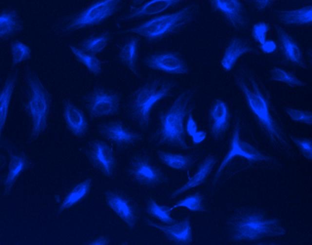
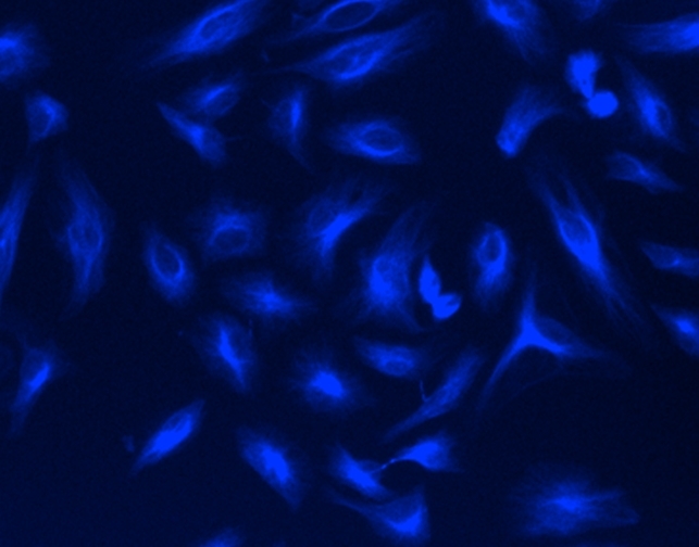
This labeling protocol was developed for the conjugate of Goat anti-mouse IgG with mFluor™ UV460 SE. You might need further optimization for your particular proteins.
Note     Each protein requires distinct dye/protein ratio, which also depends on the properties of dyes. Over labeling of a protein could detrimentally affects its binding affinity while the protein conjugates of low dye/protein ratio gives reduced sensitivity.


Run conjugation reaction
  1. Use 10:1 molar ratio of Solution B (dye)/Solution A (protein) as the starting point:  Add 5 µL of the dye stock solution (Solution B, assuming the dye stock solution is 10 mM) into the vial of the protein solution (95 µL of Solution A) with effective shaking. The concentration of the protein is ~0.05 mM assuming the protein concentration is 10 mg/mL and the molecular weight of the protein is ~200KD.
    Note     We recommend to use 10:1 molar ratio of Solution B (dye)/Solution A (protein). If it is too less or too high, determine the optimal dye/protein ratio at 5:1, 15:1 and 20:1 respectively.
  2. Continue to rotate or shake the reaction mixture at room temperature for 30-60 minutes. 

Purify the conjugation
The following protocol is an example of dye-protein conjugate purification by using a Sephadex G-25 column.
  1. Prepare Sephadex G-25 column according to the manufacture instruction.
  2. Load the reaction mixture (From "Run conjugation reaction") to the top of the Sephadex G-25 column.
  3. Add PBS (pH 7.2-7.4) as soon as the sample runs just below the top resin surface.
  4. Add more PBS (pH 7.2-7.4) to the desired sample to complete the column purification. Combine the fractions that contain the desired dye-protein conjugate.
    Note     For immediate use, the dye-protein conjugate need be diluted with staining buffer, and aliquoted for multiple uses.
    Note     For longer term storage, dye-protein conjugate solution need be concentrated or freeze dried. 

Calculators


Common stock solution preparation

Table 1. Volume of DMSO needed to reconstitute specific mass of mFluor™ UV460 SE to given concentration. Note that volume is only for preparing stock solution. Refer to sample experimental protocol for appropriate experimental/physiological buffers.

0.1 mg0.5 mg1 mg5 mg10 mg
1 mM272.287 µL1.361 mL2.723 mL13.614 mL27.229 mL
5 mM54.457 µL272.287 µL544.573 µL2.723 mL5.446 mL
10 mM27.229 µL136.143 µL272.287 µL1.361 mL2.723 mL

Molarity calculator

Enter any two values (mass, volume, concentration) to calculate the third.

Mass (Calculate)Molecular weightVolume (Calculate)Concentration (Calculate)Moles
/=x=

Spectrum


Open in Advanced Spectrum Viewer
spectrum

Spectral properties

Absorbance (nm)364
Correction Factor (260 nm)0.35
Correction Factor (280 nm)0.134
Extinction coefficient (cm -1 M -1)150001
Excitation (nm)358
Emission (nm)456
Quantum yield0.861

Product Family


NameExcitation (nm)Emission (nm)Extinction coefficient (cm -1 M -1)Quantum yieldCorrection Factor (260 nm)Correction Factor (280 nm)
mFluor™ UV375 SE3513873000010.9410.0990.138
mFluor™ UV420 SE353421750001---
mFluor™ UV455 SE3574612000010.4210.6510.406
mFluor™ UV520 SE503524800001-0.4950.518
mFluor™ UV540 SE5425609000010.3510.6340.463
mFluor™ UV610 SE5906099000010.250.9490.904

Images


Citations


View all 11 citations: Citation Explorer
Low blue carbon storage in eelgrass (Zostera marina) meadows on the Pacific Coast of Canada
Authors: Postlethwaite, V. R., McGowan, A. E., Kohfeld, K. E., Robinson, C. L. K., Pellatt, M. G.
Journal: PLoS One (2018): e0198348
Molecular characterization of DXCF cyanobacteriochromes from the cyanobacterium Acaryochloris marina identifies a blue-light power sensor
Authors: Hasegawa, M., Fushimi, K., Miyake, K., Nakajima, T., Oikawa, Y., Enomoto, G., Sato, M., Ikeuchi, M., Narikawa, R.
Journal: J Biol Chem (2018): 1713-1727
Homo- and hetero-oligomerization of hydrophobic pulmonary surfactant proteins SP-B and SP-C in surfactant phospholipid membranes
Authors: Cabre, E. J., Martinez-Calle, M., Prieto, M., Fedorov, A., Olmeda, B., Loura, L. M. S., Perez-Gil, J.
Journal: J Biol Chem (2018): 9399-9411
Simultaneous lipid and content mixing assays for in vitro reconstitution studies of synaptic vesicle fusion
Authors: Liu, X., Seven, A. B., Xu, J., Esser, V., Su, L., Ma, C., Rizo, J.
Journal: Nat Protoc (2017): 2014-2028
A biliverdin-binding cyanobacteriochrome from the chlorophyll d-bearing cyanobacterium Acaryochloris marina
Authors: Narikawa, R., Nakajima, T., Aono, Y., Fushimi, K., Enomoto, G., Ni Ni, Win, Itoh, S., Sato, M., Ikeuchi, M.
Journal: Sci Rep (2015): 7950
A new type of dual-Cys cyanobacteriochrome GAF domain found in cyanobacterium Acaryochloris marina, which has an unusual red/blue reversible photoconversion cycle
Authors: Narikawa, R., Enomoto, G., Ni Ni, Win, Fushimi, K., Ikeuchi, M.
Journal: Biochemistry (2014): 5051-9
Photosynthetic electron transport in an anoxygenic photosynthetic bacterium Afifella (Rhodopseudomonas) marina measured using PAM fluorometry
Authors: Ritchie, R. J., Runcie, J. W.
Journal: Photochem Photobiol (2013): 370-83
The slow S to M fluorescence rise in cyanobacteria is due to a state 2 to state 1 transition
Authors: Kana, R., Kotabova, E., Komarek, O., Sediva, B., Papageorgiou, G. C., Govindjee,, Prasil, O.
Journal: Biochim Biophys Acta (2012): 1237-47
Utility of a fluorescent vitamin E analogue as a probe for tocopherol transfer protein activity
Authors: Morley, S., Cross, V., Cecchini, M., Nava, P., Atkinson, J., Manor, D.
Journal: Biochemistry (2006): 1075-81
Analysis of UV-excited fluorochromes by flow cytometry using near-ultraviolet laser diodes
Authors: Telford, W. G.
Journal: Cytometry A (2004): 9-17