Products
Services
Resources
Selection Guides
About
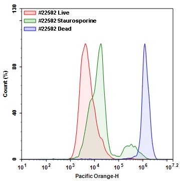
Live or Dead™ Fixable Dead Cell Staining Kit
Orange Fluorescence with 405 nm Excitation
Our Live or Dead™ Fixable Dead Cell Staining Kits are a set of tools for labeling cells for fluorescence microscopic investigations of cellular functions. The effective labeling of cells provides a powerful method for studying cellular events in a spatial and temporal context. This particular kit is designed to uniformly label fixed mammalian cells in blue fluorescence for flow cytometry applications with violet laser excitation. The kit uses a proprietary blue fluorescent dye that is more fluorescent upon bonding to cellular components. The fluorescent dye used in the kit is well excited with the violet laser (405 nm excitation) to fluorescence at 550 nm. The kit provides all the essential components with an optimized cell-labeling protocol. It is an excellent tool for preserving of fluorescent images of particular cells, and can also be used for fluorescence flow cytometry applications.
| Catalog | Size | Price | Quantity |
|---|---|---|---|
| 22502 | 200 Tests | Price |

Spectral properties
| Excitation (nm) | 394 |
| Emission (nm) | 537 |
Usage and storage
| Intended use | Research Use Only (RUO) |
Instrument settings
| Flow cytometer | |
| Excitation | 405 nm laser |
| Emission | 525/50 nm filter |
| Instrument specification(s) | Pacific Orange channel |
| Fluorescence microscope | |
| Excitation | 398 nm |
| Emission | 550 nm |
| Recommended plate | Black wall/clear bottom |
Contact us
| Telephone | |
| Fax | |
| sales@aatbio.com | |
| International | See distributors |
| Bulk request | Inquire |
| Custom size | Inquire |
| Technical Support | Contact us |
| Request quotation | Request |
| Purchase order | Send to sales@aatbio.com |
| Shipping | Standard overnight for United States, inquire for international |
Page updated on July 17, 2026