logo
AAT Bioquest

ReadiLink™ xtra Rapid Cy5 Antibody Labeling Kit *BSA-Compatible*

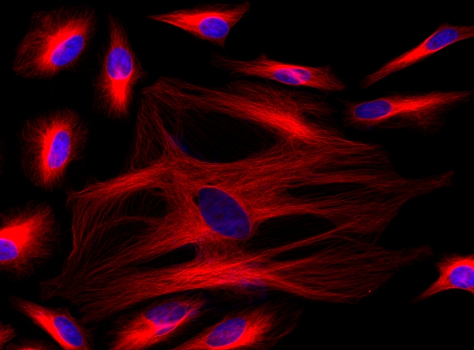
Immunofluorescence staining of tubulin in HeLa cells. HeLa cells were fixed with 4% PFA, permeabilized with 0.1% Triton X-100 and blocked. Cells were then incubated with mouse anti-tubulin monoclonal antibody and stained with a goat anti-mouse IgG labeled using the ReadiLink™ xtra Rapid Cy5 Antibody Labeling Kit (Cat No. 1972). The cell nuclei were counterstained using DAPI (Cat No. 17507).
Ordering information
Price
Catalog Number
Unit Size
Quantity
Add to cart
Additional ordering information
Telephone1-800-990-8053
Fax1-800-609-2943
Emailsales@aatbio.com
InternationalSee distributors
Bulk requestInquire
Custom sizeInquire
ShippingStandard overnight for United States, inquire for international
Request quotation
Spectral properties
Correction Factor (260 nm)0.02
Correction Factor (280 nm)0.03
Correction Factor (482 nm)0.009
Correction Factor (565 nm)0.09
Extinction coefficient (cm -1 M -1)2500001
Excitation (nm)651
Emission (nm)670
Quantum yield0.271, 0.42
Storage, safety and handling
H-phraseH303, H313, H333
Hazard symbolXN
Intended useResearch Use Only (RUO)
R-phraseR20, R21, R22
UNSPSC12171501
Related products
ReadiLink™ Rapid mFluor™ Violet 450 Antibody Labeling Kit *Microscale Optimized for Labeling 50 μg Antibody Per Reaction*
ReadiLink™ Rapid mFluor™ Violet 420 Antibody Labeling Kit *Microscale Optimized for Labeling 50 μg Antibody Per Reaction*
ReadiLink™ Rapid mFluor™ Violet 510 Antibody Labeling Kit *Microscale Optimized for Labeling 50 μg Antibody Per Reaction*
ReadiLink™ Rapid mFluor™ Violet 540 Antibody Labeling Kit *Microscale Optimized for Labeling 50 μg Antibody Per Reaction*
ReadiLink™ Rapid mFluor™ Blue 570 Antibody Labeling Kit *Microscale Optimized for Labeling 50 μg Antibody Per Reaction*
ReadiLink™ Rapid mFluor™ Green 620 Antibody Labeling Kit *Microscale Optimized for Labeling 50 μg Antibody Per Reaction*
ReadiLink™ Rapid mFluor™ Yellow 630 Antibody Labeling Kit *Microscale Optimized for Labeling 50 μg Antibody Per Reaction*
ReadiLink™ Rapid mFluor™ Red 700 Antibody Labeling Kit *Microscale Optimized for Labeling 50 μg Antibody Per Reaction*
ReadiLink™ Rapid mFluor™ Red 780 Antibody Labeling Kit *Microscale Optimized for Labeling 50 μg Antibody Per Reaction*
ReadiLink™ Rapid iFluor® 350 Antibody Labeling Kit *Microscale Optimized for Labeling 50 μg Antibody Per Reaction*
ReadiLink™ Rapid iFluor® 555 Antibody Labeling Kit *Microscale Optimized for Labeling 50 μg Antibody Per Reaction*
ReadiLink™ Rapid iFluor® 594 Antibody Labeling Kit *Microscale Optimized for Labeling 50 μg Antibody Per Reaction*
ReadiLink™ Rapid iFluor® 647 Antibody Labeling Kit *Microscale Optimized for Labeling 50 μg Antibody Per Reaction*
ReadiLink™ Rapid iFluor® 680 Antibody Labeling Kit *Microscale Optimized for Labeling 50 μg Antibody Per Reaction*
ReadiLink™ Rapid iFluor® 700 Antibody Labeling Kit *Microscale Optimized for Labeling 50 μg Antibody Per Reaction*
ReadiLink™ Rapid iFluor® 750 Antibody Labeling Kit *Microscale Optimized for Labeling 50 μg Antibody Per Reaction*
ReadiLink™ Rapid iFluor® 488 Antibody Labeling Kit *Microscale Optimized for Labeling 50 μg Antibody Per Reaction*
ReadiLink™ Rapid iFluor® 633 Antibody Labeling Kit *Microscale Optimized for Labeling 50 μg Antibody Per Reaction*
ReadiLink™ Rapid iFluor® 790 Antibody Labeling Kit *Microscale Optimized for Labeling 50 μg Antibody Per Reaction*
ReadiLink™ Rapid FITC Antibody Labeling Kit *Microscale Optimized for Labeling 50 μg Antibody Per Reaction*
ReadiLink™ Rapid trFluor™ Eu Antibody Labeling Kit *Microscale Optimized for Labeling 50 ug Antibody Per Reaction*
ReadiLink™ Rapid trFluor™ Tb Antibody Labeling Kit *Microscale Optimized for Labeling 50 ug Antibody Per Reaction*
ReadiLink™ Protein Conjugation Stop Buffer
ReadiLink™ BSA Conjugation Kit
ReadiLink™ KLH Conjugation Kit
ReadiLink™ Protein Biotinylation Kit
ReadiLink™ Protein Biotinylation Kit *Powered by ReadiView™ Biotin Visionization Technology*
ReadiLink™ Rapid Cy3 Antibody Labeling Kit *Microscale Optimized for Labeling 50 μg Antibody Per Reaction*
ReadiLink™ Rapid Cy5 Antibody Labeling Kit *Microscale Optimized for Labeling 50 μg Antibody Per Reaction*
ReadiLink™ Rapid Cy7 Antibody Labeling Kit *Microscale Optimized for Labeling 50 μg Antibody Per Reaction*
ReadiLink™ xtra Rapid iFluor® 350 Antibody Labeling Kit *BSA-Compatible*
ReadiLink™ xtra Rapid iFluor® 488 Antibody Labeling Kit *BSA-Compatible*
ReadiLink™ xtra Rapid iFluor® 555 Antibody Labeling Kit *BSA-Compatible*
ReadiLink™ xtra Rapid iFluor® 594 Antibody Labeling Kit *BSA-Compatible*
ReadiLink™ xtra Rapid iFluor® 750 Antibody Labeling Kit *BSA-Compatible*
ReadiLink™ Rapid XFD488 Antibody Labeling Kit *XFD488 Same Structure to Alexa Fluor™ 488*
ReadiLink™ Rapid XFD555 Antibody Labeling Kit *XFD555 Same Structure to Alexa Fluor™ 555*
ReadiLink™ Rapid XFD594 Antibody Labeling Kit *XFD594 Same Structure to Alexa Fluor™ 594*
ReadiLink™ Rapid XFD647 Antibody Labeling Kit *XFD647 Same Structure to Alexa Fluor™ 647*
ReadiLink™ Rapid XFD750 Antibody Labeling Kit *XFD750 Same Structure to Alexa Fluor™ 750*
ReadiLink™ iFluor® 488 Nick Translation dsDNA Labeling Kit
ReadiLink™ iFluor® 555 Nick Translation dsDNA Labeling Kit
ReadiLink™ iFluor® 647 Nick Translation dsDNA Labeling Kit
ReadiLink™ Cy3 Nick Translation dsDNA Labeling Kit
ReadiLink™ Cy5 Nick Translation dsDNA Labeling Kit
ReadiLink™ Biotin Nick Translation dsDNA Labeling Kit
ReadiLink™ DIG (Digoxigenin) Nick Translation dsDNA Labeling Kit
ReadiLink™ iFluor® 488 Oligo and ssDNA Labeling Kit
ReadiLink™ iFluor® 555 Oligo and ssDNA Labeling Kit
ReadiLink™ iFluor® 647 Oligo and ssDNA Labeling Kit
ReadiLink™ Cy3 Oligo and ssDNA Labeling Kit
ReadiLink™ Cy5 Oligo and ssDNA Labeling Kit
ReadiLink™ Biotin Oligo and ssDNA Labeling Kit
ReadiLink™ DIG (Digoxigenin) Oligo and ssDNA Labeling Kit
ReadiLink™ iFluor® 488 FISH Fluorescence Imaging Kit
ReadiLink™ iFluor® 555 FISH Fluorescence Imaging Kit
ReadiLink™ iFluor® 647 FISH Fluorescence Imaging Kit
ReadiLink™ Rapid iFluor® 350 Antibody Labeling Kit *Production Scale*
ReadiLink™ Rapid iFluor® 488 Antibody Labeling Kit *Production Scale*
ReadiLink™ Rapid iFluor® 555 Antibody Labeling Kit *Production Scale*
ReadiLink™ Rapid iFluor® 594 Antibody Labeling Kit *Production Scale*
ReadiLink™ Rapid iFluor® 647 Antibody Labeling Kit *Production Scale*
ReadiLink™ Rapid iFluor® 750 Antibody Labeling Kit *Production Scale*
ReadiLink™ Rapid FITC Antibody Labeling Kit *Production Scale*
ReadiLink™ Rapid Cy3 Antibody Labeling Kit *Production Scale*
ReadiLink™ Rapid Cy5 Antibody Labeling Kit *Production Scale*
ReadiLink™ Rapid XFD488 Antibody Labeling Kit *Production Scale*
ReadiLink™ Rapid XFD555 Antibody Labeling Kit *Production Scale*
ReadiLink™ Rapid XFD594 Antibody Labeling Kit *Production Scale*
ReadiLink™ Rapid Cy7 Antibody Labeling Kit *Production Scale*
ReadiLink™ Rapid XFD647 Antibody Labeling Kit *Production Scale*
ReadiLink™ Rapid XFD750 Antibody Labeling Kit *Production Scale*
ReadiLink™ Psoralen-A17-Biotin
ReadiLink™ Rapid mFluor™ Violet 610 Antibody Labeling Kit *Microscale Optimized for Labeling 50 µg Antibody Per Reaction*
Show More (64)

OverviewpdfSDSpdfProtocol


Correction Factor (260 nm)
0.02
Correction Factor (280 nm)
0.03
Correction Factor (482 nm)
0.009
Correction Factor (565 nm)
0.09
Extinction coefficient (cm -1 M -1)
2500001
Excitation (nm)
651
Emission (nm)
670
Quantum yield
0.271, 0.42
ReadiLink™ xtra rapid antibody labeling kits require essentially only 2 simple mixing steps without a column purification needed. Specially formulated Cy5 used in this ReadiLink™ kit is quite stable and shows good reactivity and selectivity with antibodies. The kit has all the essential components for labeling ~2x50 ug antibody. Each of the two vials of specially formulated Cy5 dye provided in the kit is optimized for labeling ~50 µg antibody. ReadiLink™ xtra Cy5 rapid antibody labeling kit provides a convenient and robust method to label monoclonal and polyclonal antibodies with the red fluorescent Cy5 fluorophore. Cy5 is one of the most used fluorophores for labeling antibodies.


readilinkworkflow


Figure 1. Overview of the ReadiLink™ xtra Rapid Antibody Labeling protocol. In just two simple steps, and with no purification necessary, covalently label microgram amounts of antibodies in under an hour.
Components


Example protocol


AT A GLANCE

Important
Warm all the components and centrifuge the vials briefly before opening, and immediately prepare the required solutions before starting your conjugation. The following protocol is for recommendation.

PREPARATION OF WORKING SOLUTION

Protein working solution (Solution A)
For labeling 50 µg of protein (assuming the target protein concentration is 1 mg/mL), mix 5 µL (10% of the total reaction volume) of Reaction Buffer (Component B) with 50 µL of the target protein solution.
Note     If you have a different protein concentration, adjust the protein volume accordingly to make ~50 µg of protein available for your labeling reaction.
Note     For labeling 100 µg of protein (assuming the target protein concentration is 1 mg/mL), mix 10 µL (10% of the total reaction volume) of Reaction Buffer (Component B) with 100 µL of the target protein solution.
Note     The protein should be dissolved in 1X phosphate buffered saline (PBS), pH 7.2 - 7.4; if the protein is dissolved in glycine buffer, it must be dialyzed against 1X PBS, pH 7.2 - 7.4, or use Amicon Ultra-0.5, Ultracel-10 Membrane, 10 kDa (cat# UFC501008 from Millipore) to remove free amines or ammonium salts (such as ammonium sulfate and ammonium acetate) that are widely used for protein precipitation.
Note     Impure antibodies or antibodies stabilized with bovine serum albumin (BSA) with 0.1 to 0.5 % will be labeled well.
Note     For optimal labeling efficiency, a final protein concentration range of 1 - 2 mg/mL is recommended, with a significantly reduced conjugation efficiency at less than 1 mg/mL.

SAMPLE EXPERIMENTAL PROTOCOL

Run conjugation reaction
  1. Add the protein working solution (Solution A) to ONE vial of labeling dye (Component A), and mix them well by repeatedly pipetting for a few times or vortex the vial for a few seconds.
    Note     If labeling 100 µg of protein, use both vials (Component A) of labeling dye by dividing the 100 µg of protein into 2 x 50 µg of protein and reacting each 50 µg of protein with one vial of labeling dye. Then combine both vials for the next step.
  2. Keep the conjugation reaction mixture at room temperature for 30 - 60 minutes.
    Note     The conjugation reaction mixture can be rotated or shaken for longer time if desired. 

Stop Conjugation reaction
  1. Add 5 µL (for 50 µg protein) or 10 µL (for 100 µg protein) which is 10% of the total reaction volume of TQ™-Dyed Quench Buffer (Component C) into the conjugation reaction mixture; mix well.
  2. Incubate at room temperature for 10 minutes. The labeled protein (antibody) is now ready to use. 

Storage of Protein Conjugate
The protein conjugate should be stored at > 0.5 mg/mL in the presence of a carrier protein (e.g., 0.1% bovine serum albumin). For longer storage, the protein conjugates could be lyophilized or divided into single-used aliquots and stored at ≤ –20 °C.

Spectrum


Open in Advanced Spectrum Viewer
spectrum

Spectral properties

Correction Factor (260 nm)0.02
Correction Factor (280 nm)0.03
Correction Factor (482 nm)0.009
Correction Factor (565 nm)0.09
Extinction coefficient (cm -1 M -1)2500001
Excitation (nm)651
Emission (nm)670
Quantum yield0.271, 0.42

Images


References


View all 50 references: Citation Explorer
Cyanine dye mediated mitochondrial targeting enhances the anti-cancer activity of small-molecule cargoes.
Authors: Nödling, Alexander R and Mills, Emily M and Li, Xuefei and Cardella, Davide and Sayers, Edward J and Wu, Shih-Hsiung and Jones, Arwyn T and Luk, Louis Y P and Tsai, Yu-Hsuan
Journal: Chemical communications (Cambridge, England) (2020): 4672-4675
Hybrid Tracers Based on Cyanine Backbones Targeting Prostate-Specific Membrane Antigen: Tuning Pharmacokinetic Properties and Exploring Dye-Protein Interaction.
Authors: Hensbergen, Albertus W and Buckle, Tessa and van Willigen, Danny M and Schottelius, Margret and Welling, Mick M and van der Wijk, Felicia A and Maurer, Tobias and van der Poel, Henk G and van der Pluijm, Gabri and van Weerden, Wytske M and Wester, Hans-Jürgen and van Leeuwen, Fijs W B
Journal: Journal of nuclear medicine : official publication, Society of Nuclear Medicine (2020): 234-241
Direct Cyanine-dCTP Labeling of DNA for Microarrays.
Authors: Rando, Oliver
Journal: Cold Spring Harbor protocols (2019)
Direct Cyanine-dUTP Labeling of RNA for Microarrays.
Authors: Rando, Oliver
Journal: Cold Spring Harbor protocols (2019)
Electrochemiluminescent aptasensor based on resonance energy transfer system between CdTe quantum dots and cyanine dyes for the sensitive detection of Ochratoxin A.
Authors: Gao, Jingwen and Chen, Ziyi and Mao, Lebao and Zhang, Wei and Wen, Wei and Zhang, Xiuhua and Wang, Shengfu
Journal: Talanta (2019): 178-183
Computational and experimental data on electrostatic density and stacking tendency of asymmetric cyanine 5 dyes.
Authors: Spa, S J and Hensbergen, A W and van der Wal, S and Kuil, J and van Leeuwen, F W B
Journal: Data in brief (2019): 50-55
Harnessing Cyanine Reactivity for Optical Imaging and Drug Delivery.
Authors: Gorka, Alexander P and Nani, Roger R and Schnermann, Martin J
Journal: Accounts of chemical research (2018): 3226-3235
Tracers for Fluorescence-Guided Surgery: How Elongation of the Polymethine Chain in Cyanine Dyes Alters the Pharmacokinetics of a Dual-Modality c[RGDyK] Tracer.
Authors: Buckle, Tessa and van Willigen, Danny M and Spa, Silvia J and Hensbergen, Albertus W and van der Wal, Steffen and de Korne, Clarize M and Welling, Mick M and van der Poel, Henk G and Hardwick, James C H and van Leeuwen, Fijs W B
Journal: Journal of nuclear medicine : official publication, Society of Nuclear Medicine (2018): 986-992
Self-Healing Organic Fluorophore of Cyanine-Conjugated Amphiphilic Polypeptide for Near-Infrared Photostable Bioimaging.
Authors: Li, Tuanwei and Liu, Le and Jing, Titao and Ruan, Zheng and Yuan, Pan and Yan, Lifeng
Journal: ACS applied materials & interfaces (2018): 14517-14530
A ruthenium(ii) complex-cyanine energy transfer scaffold based luminescence probe for ratiometric detection and imaging of mitochondrial peroxynitrite.
Authors: Zhang, Wenzhu and Liu, Yi and Gao, Quankun and Liu, Chaolong and Song, Bo and Zhang, Run and Yuan, Jingli
Journal: Chemical communications (Cambridge, England) (2018): 13698-13701