logo
AAT Bioquest

Cell Meter™ Annexin V Binding Apoptosis Assay Kit *Orange Fluorescence Optimized for Flow Cytometry*

Images of Jurkat cells in a Costar black wall/clear bottom 96-well plate stained with Cell Meter Annexin V Binding Apoptosis Assay Kit *Orange Fluorescence*. (Left): Untreated control cells. (Right): Cells treated with 1 μM staurosporine for 5 hours.
Ordering information
Price
Catalog Number
Unit Size
Quantity
Add to cart
Additional ordering information
Telephone1-800-990-8053
Fax1-800-609-2943
Emailsales@aatbio.com
InternationalSee distributors
Bulk requestInquire
Custom sizeInquire
ShippingStandard overnight for United States, inquire for international
Request quotation
Spectral properties
Correction Factor (260 nm)0.23
Correction Factor (280 nm)0.14
Extinction coefficient (cm -1 M -1)1000001
Excitation (nm)557
Emission (nm)570
Quantum yield0.641
Storage, safety and handling
H-phraseH303, H313, H333
Hazard symbolXN
Intended useResearch Use Only (RUO)
R-phraseR20, R21, R22
UNSPSC12352200
Related products
Cell Meter™ Intracellular Fluorimetric Hydrogen Peroxide Assay Kit *Green Fluorescence*
Cell Meter™ Intracellular Fluorimetric Hydrogen Peroxide Assay Kit *Blue Fluorescence*
Cell Meter™ Intracellular Fluorimetric Hydrogen Peroxide Assay Kit *Blue Fluorescence Optimized for Flow Cytometry*
Cell Meter™ Intracellular Fluorimetric Hydrogen Peroxide Assay Kit *Green Fluorescence Optimized for Flow Cytometry*
Cell Meter™ Mitochondrial Hydroxyl Radical Detection Kit *Red Fluorescence*
Cell Meter™ Fluorimetric Mitochondrial Superoxide Activity Assay Kit *Green Fluorescence*
Cell Meter™ Fluorimetric Intracellular Peroxynitrite Assay Kit *Green Fluorescence*
Cell Meter™ Fluorimetric Intracellular Peroxynitrite Assay Kit *Optimized for Flow Cytometry*
Cell Meter™ Fluorimetric Intracellular Nitric Oxide (NO) Activity Assay Kit *Orange Fluorescence Optimized for Microplate Reader*
Cell Meter™ Fluorimetric Intracellular Nitric Oxide (NO) Activity Assay Kit *Orange Fluorescence Optimized for Flow Cytometry*
Cell Meter™ Fluorimetric Intracellular Nitric Oxide (NO) Activity Assay Kit *Red Fluorescence Optimized for Flow Cytometry*
Cell Meter™ Live Cell Caspase 3/7 Binding Assay Kit *Green Fluorescence*
Cell Meter™ Live Cell Caspase 3/7 Binding Assay Kit *Red Fluorescence*
Cell Meter™ Live Cell Caspase 1 Binding Assay Kit *Green Fluorescence*
Cell Meter™ Live Cell Caspase 2 Binding Assay Kit *Green Fluorescence*
Cell Meter™ Live Cell Caspase 6 Binding Assay Kit *Green Fluorescence*
Cell Meter™ Live Cell Caspase 8 Binding Assay Kit *Green Fluorescence*
Cell Meter™ Live Cell Caspase 9 Binding Assay Kit *Green Fluorescence*
Cell Meter™ Live Cell Caspase 10 Binding Assay Kit *Green Fluorescence*
Cell Meter™ Live Cell Caspase 13 Binding Assay Kit *Green Fluorescence*
Cell Meter™ No-Wash Live Cell Caspase 3/7 Activity Assay Kit *Blue Fluorescence*
Cell Meter™ No-Wash Live Cell Caspase 3/7 Activity Assay Kit *Red Fluorescence*
Cell Meter™ Fluorimetric Intracellular pH Assay Kit
Cell Meter™ Fluorimetric Cell Cytotoxicity Assay Kit
Cell Meter™ Cell Viability Assay Kit *Red Fluorescence*
Cell Meter™ Cell Viability Assay Kit *Blue Fluorescence with 405 nm Excitation*
Cell Meter™ Cell Viability Assay Kit *Blue Fluorescence*
Cell Meter™ Cell Viability Assay Kit *Green Fluorescence*
Cell Meter™ Cell Viability Assay Kit *NIR Fluorescence Optimized for Fluorescence Microplate Reader*
Cell Meter™ Phosphatidylserine Apoptosis Assay Kit *Blue Fluorescence Optimized for Microplate Readers*
Cell Meter™ Phosphatidylserine Apoptosis Assay Kit *Green Fluorescence Optimized for Microplate Readers*
Cell Meter™ Phosphatidylserine Apoptosis Assay Kit *Red Fluorescence Optimized for Microplate Readers*
Cell Meter™ Phosphatidylserine Apoptosis Assay Kit *Deep Red Fluorescence Optimized for Microplate Readers*
Cell Meter™ Phosphatidylserine Apoptosis Assay Kit *Orange Fluorescence Optimized for Microplate Readers*
Cell Meter™ Caspase 3/7 Activity Apoptosis Assay Kit *Blue Fluorescence*
Cell Meter™ Caspase 3/7 Activity Apoptosis Assay Kit *Green Fluorescence*
Cell Meter™ Caspase 3/7 Activity Apoptosis Assay Kit *Red Fluorescence*
Cell Meter™ Caspase 8 Activity Apoptosis Assay Kit *Green Fluorescence*
Cell Meter™ Caspase 9 Activity Apoptosis Assay Kit *Green Fluorescence*
Cell Meter™ JC-10 Mitochondrion Membrane Potential Assay Kit *Optimized for Microplate Assays*
Cell Meter™ JC-10 Mitochondrion Membrane Potential Assay Kit *Optimized for Flow Cytometry Assays*
Cell Meter™ NIR Mitochondrion Membrane Potential Assay Kit *Optimized for Flow Cytometry*
Cell Meter™ NIR Mitochondrion Membrane Potential Assay Kit *Optimized for Microplate Reader*
Cell Meter™ Mitochondrion Membrane Potential Assay Kit *Orange Fluorescence Optimized for Flow Cytometry*
Cell Meter™ Mitochondrion Membrane Potential Assay Kit *Orange Fluorescence Optimized for Microplate Reader*
Cell Meter™ Intracellular GSH Assay Kit *Optimized for Flow Cytometry*
Cell Meter™ Nuclear Apoptosis Assay Kit *Green Fluorescence Optimized for Flow Cytometry*
Cell Meter™ Caspase 8 Activity Apoptosis Assay Kit *Blue Fluorescence*
Cell Meter™ Caspase 9 Activity Apoptosis Assay Kit *Blue Fluorescence*
Cell Meter™ Caspase 8 Activity Apoptosis Assay Kit *Red Fluorescence*
Cell Meter™ Caspase 9 Activity Apoptosis Assay Kit *Red Fluorescence*
Cell Meter™ Multiplexing Caspase 3/7, 8 and 9 Activity Assay Kit *Triple Fluorescence Colors*
Cell Meter™ Generic Fluorimetric Caspase Activity Assay Kit *Green Fluorescence Optimized for Flow Cytometry*
Cell Meter™ Generic Fluorimetric Caspase Binding Assay Kit *Red Fluorescence Optimized for Flow Cytometry*
Cell Meter™ Caspase 3/7 Activity Apoptosis Assay Kit *Green Fluorescence Optimized for Flow Cytometry*
Cell Meter™ Phosphatidylserine Apoptosis Assay Kit *Green Fluorescence Optimized for Flow Cytometry*
Cell Meter™ Phosphatidylserine Apoptosis Assay Kit *Deep Red Fluorescence Optimized for Flow Cytometry*
Cell Meter™ Phosphatidylserine Apoptosis Assay Kit *Blue Fluorescence Excited at 405 nm*
Cell Meter™ Phosphatidylserine Apoptosis Assay Kit *Green Fluorescence Excited at 405 nm*
Cell Meter™ APC-Annexin V Binding Apoptosis Assay Kit *Optimized for Flow Cytometry*
Cell Meter™ PE-Annexin V Binding Apoptosis Assay Kit *Optimized for Flow Cytometry*
Cell Meter™ Apoptotic and Necrotic Multiplexing Detection Kit I *Triple Fluorescence Colors*
Cell Meter™ Fluorimetric Live Cell Cycle Assay Kit *Green Fluorescence Optimized for Flow Cytometry*
Cell Meter™ Fluorimetric Fixed Cell Cycle Assay Kit *Red Fluorescence Optimized for Flow Cytometry*
Cell Meter™ Apoptotic and Necrotic Multiplexing Detection Kit II *Triple Fluorescence Colors*
Cell Meter™ Live Cell TUNEL Apoptosis Assay Kit *Red Fluorescence*
Cell Meter™ Fluorimetric Live Cell Cycle Assay Kit *Optimized for 405 nm Violet Laser Excitation*
Cell Meter™ Live Cell Caspase 3/7 and Phosphatidylserine Detection Kit *Triple Fluorescence Colors*
Cell Meter™ Fluorimetric Intracellular Total ROS Activity Assay Kit*Green Fluorescence*
Cell Meter™ Fluorimetric Intracellular Total ROS Activity Assay Kit*Red Fluorescence*
Cell Meter™ Fluorimetric Intracellular Total ROS Activity Assay Kit*Orange Fluorescence*
Cell Meter™ Fluorimetric Intracellular Total ROS Activity Assay Kit*Deep Red Fluorescence*
Cell Meter™ Fluorimetric Intracellular Total ROS Activity Assay Kit*Optimized for Flow Cytometry*
Cell Meter™ Fluorimetric Mitochondrial Superoxide Activity Assay Kit*Optimized for Flow Cytometry*
Cell Meter™ Fluorimetric Mitochondrial Superoxide Activity Assay Kit*Optimized for Microplate Reader*
Cell Meter™ Autophagy Assay Kit *Blue Fluorescence*
Cell Meter™ Autophagy Fluorescence Imaging Kit
Cell Meter™ Autophagy Assay Kit *Green Fluorescence*
Cell Meter™ FITC-Annexin V Binding Apoptosis Assay Kit *Optimized for Flow Cytometry*
Cell Meter™ Fluorimetric Intracellular Nitric Oxide (NO) Activity Assay Kit *NIR Fluorescence Optimized for Microplate Reader*
Cell Meter™ Fluorimetric Intracellular Nitric Oxide (NO) Activity Assay Kit *NIR Fluorescence Optimized for Flow Cytometry*
Cell Meter™ Colorimetric WST-8 Cell Quantification Kit
Cell Meter™ Multiplexing Live, Apoptotic and Necrotic Cell Detection Kit III *Triple Fluorescence Colors*
Cell Meter™ 2-NBDG Glucose Uptake Assay Kit
Cell Meter™ Intracellular NADH/NADPH Fluorescence Imaging Kit *Red Fluorescence*
Cell Meter™ Intracellular NADH/NADPH Flow Cytometric Analysis Kit *Red Fluorescence*
Cell Meter™ Live Cell TUNEL Apoptosis Assay Kit *Green Fluorescence*
Cell Meter™ No Wash and Probenecid-Free Endpoint Calcium Assay Kit *Optimized for microplate reader*
Cell Meter™ Mitochondrion Membrane Potential Assay Kit *Red Fluorescence Optimized for Flow Cytometry*
Cell Meter™ Mitochondrion Membrane Potential Assay Kit *Red Fluorescence Optimized for Microplate Reader*
Cell Meter™ Intracellular Colorimetric Lipid Peroxidation (MDA) Assay Kit
Cell Meter™ Flow Cytometric Calcium Assay Kit
Cell Meter™ Fluorimetric Live Cell Cycle Assay Kit *Red Fluorescence Optimized for Flow Cytometry*
Cell Meter™ Fluorimetric Cellular Lipid Peroxidation Assay Kit
Cell Meter™ Intracellular NADH/NADPH Fluorescence Imaging Kit *Deep Red Fluorescence*
Cell Meter™ Intracellular NADH/NADPH Flow Cytometric Analysis Kit *Deep Red Fluorescence*
Cell Meter™ Intracellular GSH Assay Kit *Optimized for Flow Cytometry with 405 nm excitation*
Cell Meter™ Mitochondrial Autophagy Imaging Kit *Red Fluorescence*
Cell Meter™ BX520 fixable viability dye
Cell Meter™ BX590 fixable viability dye
Cell Meter™ BX650 fixable viability dye
Cell Meter™ IX830 fixable viability dye
Cell Meter™ RX660 fixable viability dye
Cell Meter™ RX700 fixable viability dye
Cell Meter™ RX780 fixable viability dye
Cell Meter™ VX450 fixable viability dye
Cell Meter™ VX500 fixable viability dye
Cell Meter™ Live Cell Caspase 8 Binding Assay Kit *Red Fluorescence*
Cell Meter™ Fixed Cell and Tissue TUNEL Apoptosis Assay Kit *Green Fluorescence*
Cell Meter™ Fixed Cell and Tissue TUNEL Apoptosis Assay Kit *Red Fluorescence*
Cell Meter™ Fluorimetric Cellular Voltage Assay Kit
Cell Meter™ Cell Adhesion Assay Kit
Cell Meter™ Fixed Cell and Tissue TUNEL Apoptosis Assay Kit *Deep Red Fluorescence*
Cell Meter™ Fixed Cell and Tissue TUNEL Apoptosis Assay Kit *Blue Fluorescence*
Cell Meter™ Fluorescence Gap Junction Tracing Kit
Cell Meter™ Colorimetric MTT Cell Proliferation Kit
Cell Meter™ Live Cell Caspase 3/7 Imaging Kit *Green Fluorescence*
Cell Meter™ Beta-Arrestin Translocation GPCR Signaling Kit
Cell Meter™ Live Cell ATP Assay Kit
Cell Meter™ Colorimetric Cell Cytotoxicity Assay Kit
Cell Meter™ Fluorimetric Phagocytosis Assay Kit *Red Fluorescence*
Cell Meter™ Cellular Senescence Activity Assay Kit *Green Fluorescence*
Cell Meter™ Cellular Senescence Activity Assay Kit *Red Fluorescence*
Cell Meter™ Cell Proliferation Assay Kit
Cell Meter™ Glucose Uptake Imaging Kit *Red Fluorescence*
Cell Meter™ Colorimetric MTS Cell Proliferation Kit
Show More (116)

OverviewpdfSDSpdfProtocol


Correction Factor (260 nm)
0.23
Correction Factor (280 nm)
0.14
Extinction coefficient (cm -1 M -1)
1000001
Excitation (nm)
557
Emission (nm)
570
Quantum yield
0.641
Our Cell Meter™ assay kits are a set of tools for monitoring cell viability. There are a variety of parameters that can be used for monitoring cell viability. This particular kit is designed to monitor cell apoptosis through measuring the translocation of phosphatidylserine (PS). In apoptosis, PS is transferred to the outer leaflet of the plasma membrane. The appearance of phosphatidylserine on the cell surface is a universal indicator of the initial/intermediate stages of cell apoptosis and can be detected before morphological changes can be observed. This kit uses a fluorescent Annexin V that specifically binds PS. Annexin V conjugates have been demonstrated to selectively bind PS. This particular assay kit is optimized to monitor cell apoptosis using a flow cytometer with 575/26 nm emission filter set (PE channel).

Platform


Flow cytometer

Excitation488 nm or 532 nm laser
Emission575/26 nm filter
Instrument specification(s)PE channel

Components


Example protocol


AT A GLANCE

Protocol Summary
  1. Prepare cells with test compounds (200 µL/sample).

  2. Add Annexin V-iFluor® 555 assay solution.

  3. Incubate at room temperature for 30 - 60 minutes.

  4. Analyze cells using a flow cytometer with a 575/26 nm filter (PE channel) or fluorescence microscope with a Cy3 filter set.

CELL PREPARATION

For guidelines on cell sample preparation, please visit:

https://www.aatbio.com/resources/guides/cell-sample-preparation.html

SAMPLE EXPERIMENTAL PROTOCOL

Prepare and incubate cells with Annexin V-iFluor® 555
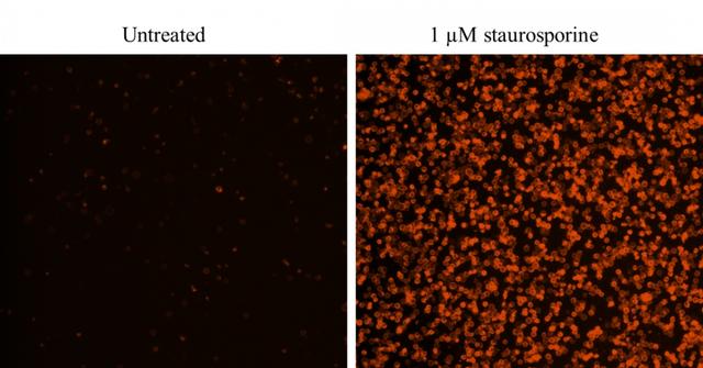
  1. Treat cells with test compounds for a desired period of time (4 - 6 hours for Jurkat cells treated with staurosporine) to induce apoptosis.

  2. Centrifuge the cells to get 1 - 5 × 10⁵ cells/tube.

  3. Resuspend cells in 200 µL of Assay Buffer (Component B).

  4. Add 2 µL of Annexin V-iFluor® 555 (Component A) into the cells.

  5. Incubate at room temperature for 30 to 60 minutes, protected from light.

  6. Add 300 µL of Assay Buffer (Component B) to increase volume before analyzing the cells with a flow cytometer or fluorescence microscope.

  7. Monitor the fluorescence intensity using a flow cytometer with a 575/26 nm filter (PE channel) or a fluorescence microscope with a Cy3 filter set.

Analyze by using a flow cytometer
  1. Quantify Annexin V- iFluor® 555 binding using a flow cytometer with a 575/26 nm filter (PE channel).

    Note: Annexin V binding flow cytometric analysis on adherent cells is not routinely tested since specific membrane damage may occur during cell detachment or harvesting. However, methods for utilizing Annexin V for flow cytometry on adherent cell types have been previously reported by Casiola-Rosen et al. and van Engelend et al.

Analyze by using a fluorescence microscope
  1. Pipette the cell suspension after incubation, rinse 1 - 2 times with Assay Buffer, and then resuspend the cells with Assay Buffer.

  2. Add the cells on a glass slide that is covered with a glass cover-slip.

    Note: For adherent cells, it is recommended to grow the cells directly on a cover-slip. After incubation with Annexin V-iFluor® 555, rinse 1 - 2 times with Assay Buffer, and add Assay Buffer back to the cover-slip. Invert the cover-slip on a glass slide and visualize the cells. The cells can also be fixed in 2% formaldehyde after incubation with Annexin V-iFluor® 555 and visualized under a microscope.

  3. Analyze the apoptotic cells with Annexin V-iFluor® 555 under a fluorescence microscope using a Cy3 filter set. Measure the cell viability by using the Cy5 channel when Nuclear Red™ DCS1 (AAT catalog #17552) is added to the cells. The orange staining on the plasma membrane indicates the Annexin V-iFluor® 555 binding to PS on the cell surface.

Spectrum


Open in Advanced Spectrum Viewer
spectrum

Spectral properties

Correction Factor (260 nm)0.23
Correction Factor (280 nm)0.14
Extinction coefficient (cm -1 M -1)1000001
Excitation (nm)557
Emission (nm)570
Quantum yield0.641

Product Family


NameExcitation (nm)Emission (nm)Extinction coefficient (cm -1 M -1)Quantum yieldCorrection Factor (260 nm)Correction Factor (280 nm)
Cell Meter™ Annexin V Binding Apoptosis Assay Kit *Green Fluorescence Optimized for Flow Cytometry*4915167500010.910.210.11
Cell Meter™ Annexin V Binding Apoptosis Assay Kit *Red Fluorescence Optimized for Flow Cytometry*58760320000010.5310.050.04
Cell Meter™ Annexin V Binding Apoptosis Assay Kit *Deep Red Fluorescence Optimized for Flow Cytometry*65667025000010.2510.030.03

Images


Citations


View all 6 citations: Citation Explorer
CCT6A knockdown suppresses osteosarcoma cell growth and Akt pathway activation in vitro
Authors: Zeng, Weiquan and Wu, Meizhu and Cheng, Ying and Liu, Liya and Han, Yuying and Xie, Qiurong and Li, Jiapeng and Wei, Lihui and Fang, Yi and Chen, Youqin and others,
Journal: Plos one (2022): e0279851
In situ polymerizable hydrogel incorporated with specific pathogen-free porcine platelet-rich plasma for the reconstruction of the corneal endothelium
Authors: Lin, Yung-Kai and Sharma, Ruchi and Ma, Hsu and Chen, Wen-Shyan and Yao, Chao-Ling
Journal: Journal of the Taiwan Institute of Chemical Engineers (2017)
Novel regulations of MEF2-A, MEF2-D, and CACNA1S in the functional incompetence of adipose-derived mesenchymal stem cells by induced indoxyl sulfate in chronic kidney disease
Authors: Do, Duyen Thi and Phan, Nam Nhut and Wang, Chih-Yang and Sun, Zhengda and Lin, Yen-Chang
Journal: Cytotechnology (2016): 2589--2604
Shear stress-induced alteration of epithelial organization in human renal tubular cells
Authors: Maggiorani, Damien and Dissard, Romain and Belloy, Marcy and Saulnier-Blache, Jean-Sébastien and Casemayou, Audrey and Ducasse, Laure and Grès, S and ra , undefined and Bellière, Julie and Caubet, Cécile and Basc, undefined and s, Jean-Loup and others, undefined
Journal: PloS one (2015): e0131416
Shear stress-induced alteration of epithelial organization in human renal tubular cells
Authors: Maggiorani, Damien and Dissard, Romain and Belloy, Marcy and Saulnier-Blache, Jean-S{\'e}bastien and Casemayou, Audrey and Ducasse, Laure and Gr{\`e}s, Sandra and Belli{\`e}re, Julie and Caubet, C{\'e}cile and Bascands, Jean-Loup and others,
Journal: PLoS One (2015): e0131416
Targeting a G-protein-coupled receptor overexpressed in endocrine tumors by magnetic nanoparticles to induce cell death
Authors: Sanchez, Claire and El Hajj Diab, Darine and Connord, Vincent and Clerc, Pascal and Meunier, Etienne and Pipy, Bernard and Payré, Bruno and Tan, Reasmey P and Gougeon, Michel and Carrey, Julian and others, undefined
Journal: Acs Nano (2014): 1350--1363

References


View all 32 references: Citation Explorer
Gold fluorescent annexin A5 as a novel apoptosis detection tool
Authors: Kurschus FC, Pal PP, Baumler P, Jenne DE, Wiltschi B, Budisa N.
Journal: Cytometry A (2009): 626
Glycogen synthase kinase-3 and Omi/HtrA2 induce annexin A2 cleavage followed by cell cycle inhibition and apoptosis
Authors: Wang CY, Lin YS, Su WC, Chen CL, Lin CF.
Journal: Mol Biol Cell (2009): 4153
Evaluation of annexin V and Calcein-AM as markers of mononuclear cell apoptosis during human immunodeficiency virus infection
Authors: Palma PF, Baggio GL, Spada C, Silva RD, Ferreira SI, Treitinger A.
Journal: Braz J Infect Dis (2008): 108
Detection of apoptosis induced by new type gosling viral enteritis virus in vitro through fluorescein annexin V-FITC/PI double labeling
Authors: Chen S, Cheng AC, Wang MS, Peng X.
Journal: World J Gastroenterol (2008): 2174
Measurement of annexin V uptake and lactadherin labeling for the quantification of apoptosis in adherent Tca8113 and ACC-2 cells
Authors: Hu T, Shi J, Jiao X, Zhou J, Yin X.
Journal: Braz J Med Biol Res (2008): 750
Two-way trafficking of Annexin V positive cells between mother and fetus: determination of apoptosis at delivery
Authors: Kolialexi A, Tsangaris GT, Anagnostopoulos A, Chondros D, Bagiokos V, Kitsiou S, Kanavakis E, Mavrou A.
Journal: Prenat Diagn (2007): 348
Rhodamine B isothiocyanate doped silica-coated fluorescent nanoparticles (RBITC-DSFNPs)-based bioprobes conjugated to Annexin V for apoptosis detection and imaging
Authors: Shi H, He X, Wang K, Yuan Y, Deng K, Chen J, Tan W.
Journal: Nanomedicine (2007): 266
Annexin A5-functionalized bimodal lipid-based contrast agents for the detection of apoptosis
Authors: van Tilborg GA, Mulder WJ, Deckers N, Storm G, Reutelingsperger CP, Strijkers GJ, Nicolay K.
Journal: Bioconjug Chem (2006): 741
Analysis of cycloheximide-induced apoptosis in human leukocytes: Fluorescence microscopy using annexin V/propidium iodide versus acridin orange/ethidium bromide
Authors: Baskic D, Popovic S, Ristic P, Arsenijevic NN.
Journal: Cell Biol Int (2006): 924
Quantum dots based probes conjugated to annexin V for photostable apoptosis detection and imaging
Authors: Le Gac S, Vermes I, van den Berg A.
Journal: Nano Lett (2006): 1863