iFluor® 555 PSA™ Imaging Kit with Goat Anti-Rabbit IgG
Example protocol
AT A GLANCE
Fix/permeabilize/block cells or tissue.
Add primary antibody in blocking buffer.
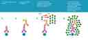
Add HRP-conjugated secondary antibody.
Prepare Styramide™ working solution and apply in cells or tissue for 5-10 minutes at room temperature.
PREPARATION OF STOCK SOLUTIONS
Unless otherwise noted, all unused stock solutions should be divided into single-use aliquots and stored at -20 °C after preparation. Avoid repeated freeze-thaw cycles
Add 100 µL of DMSO into the vial of iFluor® 555-labeled Styramide™ conjugate (Component A) to make 100X Styramide™ stock solution.
Note: Make single-use aliquots and store unused 100X stock solution at -20 °C in a dark place. Avoid repeat freeze-thaw cycles.
Add 10 µL of 3% hydrogen peroxide (Component E) to 90 µL of ddH2O.
Note: Prepare the 100X H2O2 solution fresh on the day of use.
PREPARATION OF WORKING SOLUTION
Every 1 mL of Reaction Buffer requires 10 µL of Styramide™ stock solution and 10 µL of H2O2 stock solution.
Note: The Styramide™ provided is enough for 100 tests based on 100 µL of Styramide™ working solution needed per coverslip or per well in a 96-well microplate.
Note: The Styramide™ working solution must be used within 2 hours after preparation and avoid direct exposure to light.
Dilute the 100X secondary antibody-HRP stock solution 1:100 in PBS with 1% BSA.
Note: The secondary antibody-HRP provided in this kit is sufficient for 100 tests based on 100 µL HRP working solution per coverslip or per well in a 96-well microplate.
SAMPLE EXPERIMENTAL PROTOCOL
This protocol is applicable for both cells and tissues staining.
- Fix the cells or tissue with 3.7% formaldehyde or paraformaldehyde, in PBS at room temperature for 20 minutes.
- Rinse the cells or tissue with PBS twice.
- Permeabilize the cells with 0.1% Triton X-100 solution for 1-5 minutes at room temperature.
- Rinse the cells or tissue with PBS twice.
Deparaffinize and dehydrate the tissue according to the standard IHC protocols. Perform antigen retrieval with the preferred specific solution/protocol as needed. A protocol can be found at:
https://www.aatbio.com/resources/guides/paraffin-embedded-tissue-immunohistochemistry-protocol.html
- Optional: Quench endogenous peroxidase activity by incubating cell or tissue sample in peroxidase quenching solution (such as 3% hydrogen peroxide) for 10 minutes. Rinse with PBS twice at room temperature.
- Optional: If using HRP-conjugated streptavidin, it is advisable to block endogenous biotins by biotin blocking buffer.
- Block with preferred blocking solution (such as PBS with 1% BSA) for 30 minutes at 4 °C.
- Remove blocking solution and add primary antibody diluted in recommended antibody diluent for 60 minutes at room temperature or overnight at 4 °C.
- Wash with PBS three times for 5 minutes each.
Apply 100 µL of secondary antibody-HRP working solution to each sample and incubate for 60 minutes at room temperature.
Note: Incubation time and concentration can be varied depending on the signal intensity.
- Wash with PBS three times for 5 minutes each.
Prepare and apply 100 µL of Styramide™ working solution to each sample and incubate for 5-10 minutes at room temperature.
Note: If you observe a non-specific signal, you can shorten the incubation time with Styramide. You should optimize the incubation period using positive and negative control samples at various incubation time points. Or you can use a lower concentration of Styramide in the working solution.
- Rinse with PBS three times.
Important: When performing multiplexing experiments with PSA™ Imaging Kits, it is essential to maintain a specific imaging sequence. To ensure clear and accurate results, it is recommended to image the targets from least to most abundant, with the nucleus counterstained last. Following this optimized imaging sequence will enhance the precision and quality of your experiment.
Following Step 2 in the Styramide labeling section, you can multiplex cells or tissue samples either by employing another iFluor® PSA™ Imaging Kit or by utilizing conventional IHC/ICC protocols.
For multiplexing, employ a primary antibody from a host different from the one utilized in Step 4 of the Peroxidase labeling section, along with a fluorescent label that is spectrally compatible with the initial fluorescent label.
Dilute 10X Citrate Buffer (pH 6.0) (Cat. No. 10000) 1:10 in distilled water.
Following Step 2 of the Styramide labeling process, immerse the tissue in a diluted citrate buffer (pH 6.0) and heat in a microwave oven at 100% power until boiling (1–2.5 minutes).
Reduce the power to 20% and microwave for an additional 15 minutes.
Allow the tissue sample to cool to room temperature while it remains in the citrate buffer.
Wash the sample twice with 1X PBS. Then, repeat the Peroxidase labeling and Styramide labeling steps using a primary antibody of the same species, if desired.
Use an iFluor® PSA™ Imaging Kit that is spectrally compatible with the iFluor® 350 PSA™ Imaging Kit used in the first round.
Counterstain the cell or tissue samples as needed. AAT provides a series of nucleus counterstain reagents as listed in Table 1. Follow the instructions provided with the reagents.
Mount the coverslip using a mounting medium with anti-fading properties.
Note: To ensure optimal results, it is recommended to use either ReadiUse™ microscope mounting solution (Cat. 20009) or FluoroQuest™ TSA/PSA Antifade Mounting Medium *Optimized for Tyramide and Styramide Imaging* (Cat. 44890) instead of Vectashield® mounting media. There are instances where Vectashield® mounting media may not be suitable for certain TSA/PSA conjugates.
Use the appropriate filter set to visualize the signal from the Styramide labeling.
Table 1. Recommended nucleus counterstains.
Cat# | Product Name | Ex/Em (nm) |
17548 | Nuclear Blue™ DCS1 | 350/461 |
17550 | Nuclear Green™ DCS1 | 503/526 |
17551 | Nuclear Orange™ DCS1 | 528/576 |
17552 | Nuclear Red™ DCS1 | 642/660 |
Spectrum

Product family
| Name | Excitation (nm) | Emission (nm) | Extinction coefficient (cm -1 M -1) | Quantum yield | Correction Factor (260 nm) | Correction Factor (280 nm) |
| iFluor® 350 PSA™ Imaging Kit with Goat Anti-Rabbit IgG | 345 | 450 | 200001 | 0.951 | 0.83 | 0.23 |
| iFluor® 488 PSA™ Imaging Kit with Goat Anti-Rabbit IgG | 491 | 516 | 750001 | 0.91 | 0.21 | 0.11 |
| iFluor® 555 PSA™ Imaging Kit with Goat Anti-Mouse IgG | 557 | 570 | 1000001 | 0.641 | 0.23 | 0.14 |
| iFluor® 594 PSA™ Imaging Kit with Goat Anti-Rabbit IgG | 587 | 603 | 2000001 | 0.531 | 0.05 | 0.04 |
| iFluor® 647 PSA™ Imaging Kit with Goat Anti-Rabbit IgG | 656 | 670 | 2500001 | 0.251 | 0.03 | 0.03 |