Products
Services
Resources
Selection Guides
About
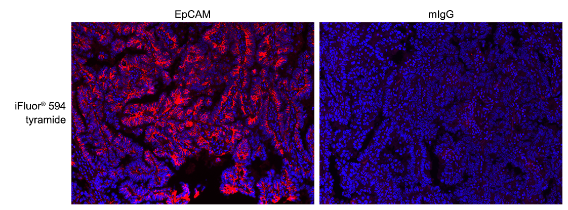
iFluor® 594 Tyramide
For many immunohistochemical (IHC) applications, traditional enzymatic amplification procedures are sufficient for achieving adequate antigen detection. However, several factors limit their sensitivity and utility. Tyramide signal amplification (TSA) has proven to be a particularly versatile and powerful enzyme amplification technique with improved assay sensitivity. TSA is based on the ability of HRP, in the presence of low concentrations of hydrogen peroxide, to convert labeled tyramine-containing substrate into an oxidized, highly reactive free radical that can covalently bind to tyrosine residues at or near the HRP. To achieve maximal IHC detection, tyramine is prelabeled with a fluorophore. The signal amplification conferred by the turnover of multiple tyramide substrates per peroxidase label results in the ability to detect low-abundance targets with ultrasensitive precision and reduces the amount of antibodies and hybridization probes needed. In IHC applications, this method can also enhance sensitivity in cases where the primary antibody dilution needs to be increased to reduce nonspecific background signals or overcome weak immunolabeling due to suboptimal fixation procedures or low levels of target expression. The iFluor® 594 tyramide contains the bright iFluor® 594 that can be readily detected with the standard Texas Red filter set, as well as with a TRITC or Cy3 filter set. iFluor® dyes have higher fluorescence intensity, increased photostability, and enhanced water solubility, resulting in fluorescence signals with significantly higher precision and sensitivity. iFluor® 594 is an excellent replacement for Alexa Fluor® 594 tyramide (Alexa Fluor® is the trademark of ThermoFisher) or other comparable fluorescent tyramide conjugates.
| Catalog | Size | Price | Quantity |
|---|---|---|---|
| 45107 | 200 Slides | Price |

Physical properties
| Molecular weight | 841.95 |
| Solvent | DMSO |
Spectral properties
| Absorbance (nm) | 587 |
| Correction factor (260 nm) | 0.05 |
| Correction factor (280 nm) | 0.04 |
| Extinction coefficient (cm -1 M -1) | 200000 1 |
| Excitation (nm) | 587 |
| Emission (nm) | 603 |
| Quantum yield | 0.53 1 |
Usage and storage
| Intended use | Research Use Only (RUO) |
| Storage | Freeze (< -15 °C); Minimize light exposure |
Instrument settings
| Fluorescence microscope | |
| Excitation | Cy3/TRITC filter set |
| Emission | Cy3/TRITC filter set |
| Recommended plate | Black wall/clear bottom |
| Instrument specification(s) | Compatible with Texas Red filter set |
Contact us
| Telephone | |
| Fax | |
| sales@aatbio.com | |
| International | See distributors |
| Bulk request | Inquire |
| Custom size | Inquire |
| Technical Support | Contact us |
| Request quotation | Request |
| Purchase order | Send to sales@aatbio.com |
| Shipping | Standard overnight for United States, inquire for international |
Page updated on July 26, 2026