iFluor® 430 Tyramide *Superior Replacement for Opal 480*
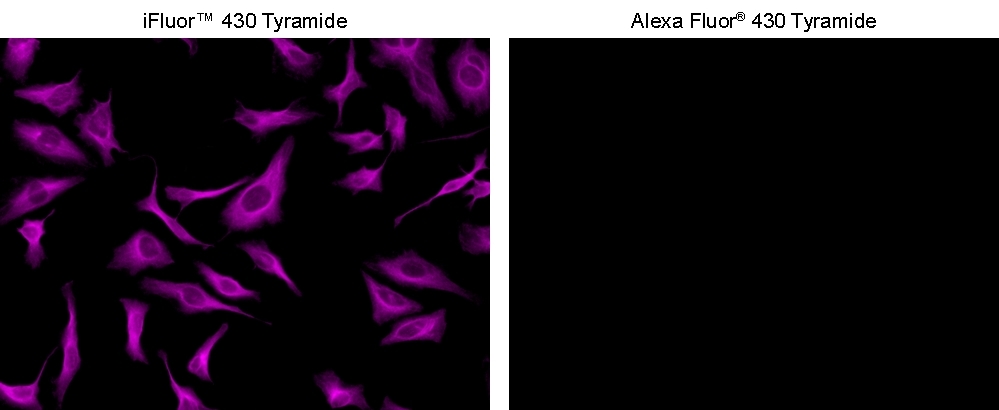
iFluor® 430 tyramide is optimized to a superior replacement for Opal 480 or other spectrally similar fluorescent tyramide conjugates or TSA reagents. Tyramide reagetns can be used to detect extremely low-abundance targets in cells and tissues with significantly improved fluorescence signal than the direct fluorescence labeling reagents. In combination with our superior iFluor® dyes that have higher florescence intensity, increased photostability and enhanced water solubility, the iFluor® dye-labeled tyramide conjugates can generate fluorescence signal with significantly higher precision and sensitivity.
Example protocol
AT A GLANCE
Protocol Summary
- Fix/permeabilize/block cells or tissue
- Add primary antibody in blocking buffer
- Add HRP-conjugated secondary antibody
- Prepare tyramide working solution and apply in cells or tissue for 5-10 minutes at room temperature
PREPARATION OF STOCK SOLUTIONS
Unless otherwise noted, all unused stock solutions should be divided into single-use aliquots and stored at -20 °C after preparation. Avoid repeated freeze-thaw cycles.
Note Unused tyramide stock solution can be stored at 2-8 °C.
iFluor™ 430 Tyramide stock solution (200X)
Add 100 µL DMSO to vial and mix well.Note Unused tyramide stock solution can be stored at 2-8 °C.
PREPARATION OF WORKING SOLUTION
iFluor™ 430 Tyramide working solution (1X)
Add 100 µL of Tyramide stock solution into 20 mL of buffer of your choice containing 0.003% H2O2.Note Tris Buffer, pH=7.4 can be used for optimal performance.
Note Tyramide working solution should be used immediately and made fresh on the day of use.
Note 20 mL solution is good for 200 tests.
SAMPLE EXPERIMENTAL PROTOCOL
This protocol is applicable for both cells and tissues staining.
Protocol can be found at
https://www.aatbio.com/resources/guides/paraffin-embedded-tissue-immunohistochemistry-protocol.html
Cell fixation and permeabilization
- Fix the cells or tissue with 3.7% formaldehyde or paraformaldehyde, in PBS at room temperature for 20 minutes.
- Rinse the cells or tissue with PBS twice.
- Permeabilize the cells with 0.1% Triton X-100 solution for 1-5 minutes at room temperature.
- Rinse the cells or tissue with PBS twice.
Tissue fixation, deparaffinization and rehydration
Deparaffinize and dehydrate the tissue according to the standard IHC protocols. Perform antigen retrieval with preferred specific solution/protocol as needed.Protocol can be found at
https://www.aatbio.com/resources/guides/paraffin-embedded-tissue-immunohistochemistry-protocol.html
Peroxidase labeling
- Optional: Quench endogenous peroxidase activity by incubating cell or tissue sample in peroxidase quenching solution (such as 3% hydrogen peroxide) for 10 minutes. Rinse with PBS twice at room temperature.
- Optional: If using HRP-conjugated streptavidin, it is advisable to block endogenous biotins by biotin blocking buffer.
- Block with preferred blocking solution (such as PBS with 1% BSA) for 30 minutes at 4 °C.
- Remove blocking solution and add primary antibody diluted in recommended antibody diluent for 60 minutes at room temperature or overnight at 4 °C.
- Wash with PBS three times for 5 minutes each.
- Apply 100 µL of secondary antibody-HRP working solution to each sample and incubate for 60 minutes at room temperature.
Note Incubation time and concentration can be varied depending on the signal intensity. - Wash with PBS three times for 5 minutes each.
Tyramide labeling
- Prepare and apply 100 µL of tyramide working solution to each sample and incubate for 5-10 minutes at room temperature.
Note If you observe non-specific signal, you can shorten the incubation time with tyramide. You should optimize the incubation period using positive and negative control samples at various incubation time points. Or you can use lower concentration of tyramide in the working solution. - Rinse with PBS three times.
Counterstain and fluorescence imaging
- Counterstain the cell or tissue samples as needed. AAT provides a series of nucleus counterstain reagents as listed in Table 1. Follow the instruction provided with the reagents.
- Mount the coverslip using a mounting medium with anti-fading properties.
- Use the appropriate filter set to visualize the signal from the tyramide labeling.
Cat# | Product Name | Ex/Em (nm) |
17548 | Nuclear Blue™ DCS1 | 350/461 |
17550 | Nuclear Green™ DCS1 | 503/526 |
17551 | Nuclear Orange™ DCS1 | 528/576 |
17552 | Nuclear Red™ DCS1 | 642/660 |
Spectrum
Open in Advanced Spectrum Viewer
Product family
Name | Excitation (nm) | Emission (nm) | Extinction coefficient (cm -1 M -1) | Quantum yield | Correction Factor (260 nm) | Correction Factor (280 nm) |
iFluor® 488 tyramide | 491 | 516 | 750001 | 0.91 | 0.21 | 0.11 |
iFluor® 430 maleimide | 433 | 498 | 400001 | 0.781 | 0.68 | 0.3 |
iFluor® 555 Tyramide | 557 | 570 | 1000001 | 0.641 | 0.23 | 0.14 |
iFluor® 647 Tyramide | 656 | 670 | 2500001 | 0.251 | 0.03 | 0.03 |
iFluor® 350 Tyramide | 345 | 450 | 200001 | 0.951 | 0.83 | 0.23 |
iFluor® 546 Tyramide | 541 | 557 | 1000001 | 0.671 | 0.25 | 0.15 |
iFluor® 568 Tyramide | 568 | 587 | 1000001 | 0.571 | 0.34 | 0.15 |
iFluor® 594 Tyramide | 587 | 603 | 2000001 | 0.531 | 0.05 | 0.04 |
iFluor® 633 tyramide | 640 | 654 | 2500001 | 0.291 | 0.062 | 0.044 |
Show More (3) |
Citations
View all 2 citations: Citation Explorer
Intratumoral injection of interferon gamma promotes the efficacy anti-PD1 treatment in colorectal cancer
Authors: Tang, Yang and Wei, Jingsun and Ge, Xiaoxu and Yu, Chengxuan and Lu, Wei and Qian, Yucheng and Yang, Hang and Fu, Dongliang and Fang, Yimin and Zhou, Yinyi and others,
Journal: Cancer Letters (2024): 216798
Authors: Tang, Yang and Wei, Jingsun and Ge, Xiaoxu and Yu, Chengxuan and Lu, Wei and Qian, Yucheng and Yang, Hang and Fu, Dongliang and Fang, Yimin and Zhou, Yinyi and others,
Journal: Cancer Letters (2024): 216798
MDIG-mediated H3K9me3 demethylation upregulates Myc by activating OTX2 and facilitates liver regeneration
Authors: Du, Jinpeng and Liao, Wenwei and Wang, Haichuan and Hou, Guimin and Liao, Min and Xu, Lin and Huang, Jiwei and Yuan, Kefei and Chen, Xiangzheng and Zeng, Yong
Journal: Signal Transduction and Targeted Therapy (2023): 351
Authors: Du, Jinpeng and Liao, Wenwei and Wang, Haichuan and Hou, Guimin and Liao, Min and Xu, Lin and Huang, Jiwei and Yuan, Kefei and Chen, Xiangzheng and Zeng, Yong
Journal: Signal Transduction and Targeted Therapy (2023): 351
References
View all 50 references: Citation Explorer
Immunofluorescent Staining of Adult Murine Paraffin-Embedded Skeletal Tissue.
Authors: Felsenthal, Neta and Zelzer, Elazar
Journal: Methods in molecular biology (Clifton, N.J.) (2021): 337-344
Authors: Felsenthal, Neta and Zelzer, Elazar
Journal: Methods in molecular biology (Clifton, N.J.) (2021): 337-344
Highly Sensitive and Multiplexed In Situ RNA Profiling with Cleavable Fluorescent Tyramide.
Authors: Xiao, Lu and Labaer, Joshua and Guo, Jia
Journal: Cells (2021)
Authors: Xiao, Lu and Labaer, Joshua and Guo, Jia
Journal: Cells (2021)
Single-cell RNA sequencing of human liver reveals hepatic stellate cell heterogeneity.
Authors: Payen, Valéry L and Lavergne, Arnaud and Alevra Sarika, Niki and Colonval, Megan and Karim, Latifa and Deckers, Manon and Najimi, Mustapha and Coppieters, Wouter and Charloteaux, Benoît and Sokal, Etienne M and El Taghdouini, Adil
Journal: JHEP reports : innovation in hepatology (2021): 100278
Multiplexed In Situ Protein Profiling with High-Performance Cleavable Fluorescent Tyramide.
Authors: Pham, Thai and Liao, Renjie and Labaer, Joshua and Guo, Jia
Journal: Molecules (Basel, Switzerland) (2021)
Authors: Pham, Thai and Liao, Renjie and Labaer, Joshua and Guo, Jia
Journal: Molecules (Basel, Switzerland) (2021)
Accessibility-dependent topology studies of membrane proteins using a SpyTag/SpyCatcher protein-ligation system.
Authors: Bae, Yoonji and Lee, Sang Kwon and Chae, Young Chan and Park, Chan Young and Kang, Sebyung
Journal: International journal of biological macromolecules (2021): 171-178
Authors: Bae, Yoonji and Lee, Sang Kwon and Chae, Young Chan and Park, Chan Young and Kang, Sebyung
Journal: International journal of biological macromolecules (2021): 171-178
Page updated on September 15, 2024