iFluor® 350 Tyramide

| Price | |
| Catalog Number | |
| Unit Size | |
| Quantity |
| Telephone | 1-800-990-8053 |
| Fax | 1-800-609-2943 |
| sales@aatbio.com | |
| International | See distributors |
| Bulk request | Inquire |
| Custom size | Inquire |
| Shipping | Standard overnight for United States, inquire for international |
| Molecular weight | 432.45 |
| Solvent | DMSO |
| Correction Factor (260 nm) | 0.83 |
| Correction Factor (280 nm) | 0.23 |
| Extinction coefficient (cm -1 M -1) | 200001 |
| Excitation (nm) | 345 |
| Emission (nm) | 450 |
| Quantum yield | 0.951 |
| H-phrase | H303, H313, H333 |
| Hazard symbol | XN |
| Intended use | Research Use Only (RUO) |
| R-phrase | R20, R21, R22 |
| Storage | Freeze (< -15 °C); Minimize light exposure |
| UNSPSC | 12171501 |
| Overview |
Molecular weight 432.45 | Correction Factor (260 nm) 0.83 | Correction Factor (280 nm) 0.23 | Extinction coefficient (cm -1 M -1) 200001 | Excitation (nm) 345 | Emission (nm) 450 | Quantum yield 0.951 |
Platform
Fluorescence microscope
| Excitation | DAPI filter set |
| Emission | DAPI filter set |
| Recommended plate | Black wall/clear bottom |
Example protocol
AT A GLANCE
- Fix/permeabilize/block cells or tissue
- Add primary antibody in blocking buffer
- Add HRP-conjugated secondary antibody
- Prepare tyramide working solution and apply in cells or tissue for 5-10 minutes at room temperature
PREPARATION OF STOCK SOLUTIONS
Unless otherwise noted, all unused stock solutions should be divided into single-use aliquots and stored at -20 °C after preparation. Avoid repeated freeze-thaw cycles
Add 100 µL of DMSO to the vial of iFluor® tyramide and mix well.
Note: Make single-use aliquots and store unused 200X stock solution at 2-8 °C, protected from light. Avoid repeat freeze-thaw cycles.
PREPARATION OF WORKING SOLUTION
Add 100 µL of the tyramide stock solution into 20 mL of a buffer of your choice containing 0.003% H2O2.
Note: For optimal performance use Tris Buffer, pH=7.4.
Note: A 20 mL solution is good for 200 tests. The tyramide working solution should be used immediately and made fresh on the day of use. Avoid direct exposure to light.
Make an appropriate concentration of secondary antibody-HRP working solution as per the manufacturer's recommendations.
SAMPLE EXPERIMENTAL PROTOCOL
This protocol is applicable for both cells and tissues staining.
Fix the cells or tissue with 3.7% formaldehyde or paraformaldehyde, in PBS at room temperature for 20 minutes.
Rinse the cells or tissue with PBS twice.
Permeabilize the cells with 0.1% Triton X-100 solution for 1-5 minutes at room temperature.
Rinse the cells or tissue with PBS twice.
Deparaffinize and dehydrate the tissue according to the standard IHC protocols. Perform antigen retrieval with preferred specific solution/protocol as needed. A protocol can be found at:
https://www.aatbio.com/resources/guides/paraffin-embedded-tissue-immunohistochemistry-protocol.html
Optional: Quench endogenous peroxidase activity by incubating cell or tissue sample in peroxidase quenching solution (such as 3% hydrogen peroxide) for 10 minutes. Rinse with PBS twice at room temperature.
Optional: If using HRP-conjugated streptavidin, it is advisable to block endogenous biotins using a biotin blocking buffer.
Block with preferred blocking solution (such as PBS with 1% BSA) for 30 minutes at 4 °C.
Remove blocking solution and add primary antibody diluted in recommended antibody diluent for 60 minutes at room temperature or overnight at 4 °C.
Wash with PBS three times for 5 minutes each.
Apply 100 µL of secondary antibody-HRP working solution to each sample and incubate for 60 minutes at room temperature.
Note: Incubation time and concentration can be varied depending on the signal intensity.
Wash with PBS three times for 5 minutes each.
Prepare and apply 100 µL of Tyramide working solution to each sample and incubate for 5-10 minutes at room temperature.
Note: If you observe non-specific signal, you can shorten the incubation time with Tyramide. You should optimize the incubation period using positive and negative control samples at various incubation time points. Or you can use lower concentration of Tyramide in the working solution.
Rinse with PBS three times.
Counterstain the cell or tissue samples as needed. AAT provides a series of nucleus counterstain reagents as listed in Table 1. Follow the instruction provided with the reagents.
Mount the coverslip using a mounting medium with anti-fading properties.
Note: To ensure optimal results, it is recommended to use either ReadiUse™ microscope mounting solution (Cat. 20009) or FluoroQuest™ TSA/PSA Antifade Mounting Medium *Optimized for Tyramide and Styramide Imaging* (Cat. 44890) instead of Vectashield® mounting media. There are instances where Vectashield® mounting media may not be suitable for certain TSA/PSA conjugates.
Use the appropriate filter set to visualize the signal from the Tyramide labeling.
Table 1. Products recommended for nucleus counterstaining.
Cat# | Product Name | Ex/Em (nm) |
17548 | Nuclear Blue™ DCS1 | 350/461 |
17550 | Nuclear Green™ DCS1 | 503/526 |
17551 | Nuclear Orange™ DCS1 | 528/576 |
17552 | Nuclear Red™ DCS1 | 642/660 |
Calculators
Common stock solution preparation
| 0.1 mg | 0.5 mg | 1 mg | 5 mg | 10 mg | |
| 1 mM | 231.241 µL | 1.156 mL | 2.312 mL | 11.562 mL | 23.124 mL |
| 5 mM | 46.248 µL | 231.241 µL | 462.481 µL | 2.312 mL | 4.625 mL |
| 10 mM | 23.124 µL | 115.62 µL | 231.241 µL | 1.156 mL | 2.312 mL |
Molarity calculator
| Mass (Calculate) | Molecular weight | Volume (Calculate) | Concentration (Calculate) | Moles | ||||
| / | = | x | = |
Spectrum
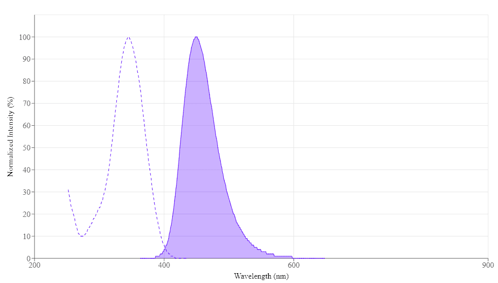
Spectral properties
| Correction Factor (260 nm) | 0.83 |
| Correction Factor (280 nm) | 0.23 |
| Extinction coefficient (cm -1 M -1) | 200001 |
| Excitation (nm) | 345 |
| Emission (nm) | 450 |
| Quantum yield | 0.951 |
Product Family
| Name | Excitation (nm) | Emission (nm) | Extinction coefficient (cm -1 M -1) | Quantum yield | Correction Factor (260 nm) | Correction Factor (280 nm) |
| iFluor® 350 maleimide | 345 | 450 | 200001 | 0.951 | 0.83 | 0.23 |
| iFluor® 350 amine | 345 | 450 | 200001 | 0.951 | 0.83 | 0.23 |
| iFluor® 350 hydrazide | 345 | 450 | 200001 | 0.951 | 0.83 | 0.23 |
| iFluor® 488 tyramide | 491 | 516 | 750001 | 0.91 | 0.21 | 0.11 |
| iFluor® 350 Styramide *Superior Replacement for Alexa Fluor 350 tyramide* | 345 | 450 | 200001 | 0.951 | 0.83 | 0.23 |
| iFluor® 555 Tyramide | 557 | 570 | 1000001 | 0.641 | 0.23 | 0.14 |
| iFluor® 647 Tyramide | 656 | 670 | 2500001 | 0.251 | 0.03 | 0.03 |
| iFluor® 546 Tyramide | 541 | 557 | 1000001 | 0.671 | 0.25 | 0.15 |
| iFluor® 568 Tyramide | 568 | 587 | 1000001 | 0.571 | 0.34 | 0.15 |
Show More (5) | ||||||
Images
References
Authors: Kuwajima, Masaaki and Ostrovskaya, Olga I and Cao, Guan and Weisberg, Seth A and Harris, Kristen M and Zemelman, Boris V
Journal: PloS one (2020): e0226797
Authors: Yamazaki, Masaki and Kato, Atsuhiko and Zaitsu, Yoko and Watanabe, Takeshi and Iimori, Makoto and Funahashi, Shinichi and Kitao, Hiroyuki and Saeki, Hiroshi and Oki, Eiji and Suzuki, Masami
Journal: Acta histochemica et cytochemica (2015): 159-64
Authors: Clutter, Matthew R and Heffner, Garrett C and Krutzik, Peter O and Sachen, Kacey L and Nolan, Garry P
Journal: Cytometry. Part A : the journal of the International Society for Analytical Cytology (2010): 1020-31
Authors: Symonds, Daniel A and Merchenthaler, Istvan and Flaws, Jodi A
Journal: Toxicological sciences : an official journal of the Society of Toxicology (2008): 182-7
Authors: Tubbs, Raymond R and Das, Kingshuk and Cook, James R and Pettay, James D and Roche, Patrick C and Grogan, Thomas
Journal: Journal of molecular histology (2007): 129-34
Authors: Tujula, Niina A and Holmström, Carola and Mussmann, Marc and Amann, Rudolf and Kjelleberg, Staffan and Crocetti, Gregory R
Journal: Journal of microbiological methods (2006): 604-7
Authors: Krieg, Reimar and Halbhuber, Karl-Jürgen
Journal: Journal of molecular histology (2004): 471-87
Authors: Warsen, Adelaide E and Krug, Melissa J and LaFrentz, Stacey and Stanek, Danielle R and Loge, Frank J and Call, Douglas R
Journal: Applied and environmental microbiology (2004): 4216-21
Authors: Panicker, Gitika and Call, Douglas R and Krug, Melissa J and Bej, Asim K
Journal: Applied and environmental microbiology (2004): 7436-44
Authors: Call, Douglas R and Bakko, Marlene K and Krug, Melissa J and Roberts, Marilyn C
Journal: Antimicrobial agents and chemotherapy (2003): 3290-5
Application notes
A Novel Fluorescent Probe for Imaging and Detecting Hydroxyl Radical in Living Cells
A Novel NO Wash Probeniceid-Free Calcium Assay for Functional Analysis of GPCR and Calcium Channel Targets
Biotin Labeling Molecules and Their Biological Applications
Buccutite™ Bioconjugation Technology
FAQ
What are the differences between calcium ion indicators: Cal 520, Cal 520FF, and Cal 520N?
How do I make an AM ester stock solution?
Can we fix cells with glutaraldehyde and then stain with fluorescent phalloidin?
What is the difference between FluoroQuest Anti-fading Kit I and FluoroQuest Anti-fading Kit II?