logo
AAT Bioquest

iFluor® 488 tyramide

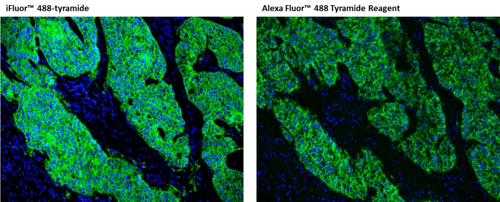
Immunofluorescent image of paraffin-embedded human lung carcinoma labeled with  EpCAM Rabbit mAb followed with HRP-labeled goat anti-rabbit IgG (H+L) (Cat#16793) . The signal was developed with iFluor® 488 tyramide or Alexa Fluor™ 488 Tyramide Reagent (Green). Cells were also counterstained with DAPI (Blue).
Immunofluorescent image of paraffin-embedded human lung carcinoma labeled with Pan-Keratin Mouse mAb followed with HRP-labeled goat anti-mouse IgG (H+L) (Cat#16728). The signal was developed with iFluor® 488 tyramide (Green). Cells were also counterstained with DAPI (Blue).
<strong>Superior sensitivity with iFluor® 488 tyramide.</strong> HeLa cells were incubated with primary anti-tubulin antibodies followed by detection with HRP-Goat anti-Mouse&nbsp;IgG and<strong><em>&nbsp;</em></strong>iFluor® 488 tyramide (Left) or Alexa Fluor&reg; 488 tyramide (Right). Fluorescence images were taken on a Keyence BZ-X710 fluorescence microscope equipped with a FITC filter set.
Formalin-fixed, paraffin-embedded (FFPE) human lung adenocarcinoma tissue was incubated with an anti-EpCAM primary antibody, and an HRP conjugated anti-mouse secondary antibody. TSA signal was developed by incubation of tissue section with 5 µg/mL of iFluor® 488 tyramide (Cat No. 45100) for 10 minutes. Images were acquired on a confocal microscope equipped with a GFP filter set.
Ordering information
Price
Catalog Number
Unit Size
Quantity
Add to cart
Additional ordering information
Telephone1-800-990-8053
Fax1-800-609-2943
Emailsales@aatbio.com
InternationalSee distributors
Bulk requestInquire
Custom sizeInquire
ShippingStandard overnight for United States, inquire for international
Request quotation
Physical properties
Molecular weight531.47
SolventDMSO
Spectral properties
Correction Factor (260 nm)0.21
Correction Factor (280 nm)0.11
Extinction coefficient (cm -1 M -1)750001
Excitation (nm)491
Emission (nm)516
Quantum yield0.91
Storage, safety and handling
H-phraseH303, H313, H333
Hazard symbolXN
Intended useResearch Use Only (RUO)
R-phraseR20, R21, R22
StorageFreeze (< -15 °C); Minimize light exposure
UNSPSC12352200
Related products
iFluor® A7 SE
iFluor® 350 maleimide
iFluor® 555 maleimide
iFluor® 647 maleimide
iFluor® 680 maleimide
iFluor® 700 maleimide
iFluor® 750 maleimide
iFluor® 350 amine
iFluor® 405 amine
iFluor® 555 amine
iFluor® 647 amine
iFluor® 660 amine
iFluor® 680 amine
iFluor® 700 amine
iFluor® 710 amine
iFluor® 750 amine
iFluor® 350 hydrazide
iFluor® 555 hydrazide
iFluor® 647 hydrazide
iFluor® 680 hydrazide
iFluor® 700 hydrazide
iFluor® 750 hydrazide
iFluor® 647 alkyne
iFluor® 647 azide
iFluor® 790 acid
iFluor® 790 amine
iFluor® 790 hydrazide
iFluor® 790 maleimide
iFluor® 350 goat anti-mouse IgG (H+L)
iFluor® 405 goat anti-mouse IgG (H+L)
iFluor® 488 goat anti-mouse IgG (H+L)
iFluor® 514 goat anti-mouse IgG (H+L)
iFluor® 532 goat anti-mouse IgG (H+L)
iFluor® 555 goat anti-mouse IgG (H+L)
iFluor® 594 goat anti-mouse IgG (H+L)
iFluor® 633 goat anti-mouse IgG (H+L)
iFluor® 647 goat anti-mouse IgG (H+L)
iFluor® 680 goat anti-mouse IgG (H+L)
iFluor® 700 goat anti-mouse IgG (H+L)
iFluor® 750 goat anti-mouse IgG (H+L)
iFluor® 790 goat anti-mouse IgG (H+L)
iFluor® 350 goat anti-mouse IgG (H+L) *Cross Adsorbed*
iFluor® 405 goat anti-mouse IgG (H+L) *Cross Adsorbed*
iFluor® 488 goat anti-mouse IgG (H+L) *Cross Adsorbed*
iFluor® 514 goat anti-mouse IgG (H+L) *Cross Adsorbed*
iFluor® 532 goat anti-mouse IgG (H+L) *Cross Adsorbed*
iFluor® 555 goat anti-mouse IgG (H+L) *Cross Adsorbed*
iFluor® 594 goat anti-mouse IgG (H+L) *Cross Adsorbed*
iFluor® 633 goat anti-mouse IgG (H+L) *Cross Adsorbed*
iFluor® 647 goat anti-mouse IgG (H+L) *Cross Adsorbed*
iFluor® 680 goat anti-mouse IgG (H+L) *Cross Adsorbed*
iFluor® 700 goat anti-mouse IgG (H+L) *Cross Adsorbed*
iFluor® 750 goat anti-mouse IgG (H+L) *Cross Adsorbed*
iFluor® 790 goat anti-mouse IgG (H+L) *Cross Adsorbed*
iFluor® 350 goat anti-rabbit IgG (H+L)
iFluor® 405 goat anti-rabbit IgG (H+L)
iFluor® 488 goat anti-rabbit IgG (H+L)
iFluor® 514 goat anti-rabbit IgG (H+L)
iFluor® 532 goat anti-rabbit IgG (H+L)
iFluor® 555 goat anti-rabbit IgG (H+L)
iFluor® 594 goat anti-rabbit IgG (H+L)
iFluor® 633 goat anti-rabbit IgG (H+L)
iFluor® 647 goat anti-rabbit IgG (H+L)
iFluor® 680 goat anti-rabbit IgG (H+L)
iFluor® 700 goat anti-rabbit IgG (H+L)
iFluor® 750 goat anti-rabbit IgG (H+L)
iFluor® 790 goat anti-rabbit IgG (H+L)
iFluor® 350 goat anti-rabbit IgG (H+L) *Cross Adsorbed*
iFluor® 405 goat anti-rabbit IgG (H+L) *Cross Adsorbed*
iFluor® 488 goat anti-rabbit IgG (H+L) *Cross Adsorbed*
iFluor® 514 goat anti-rabbit IgG (H+L) *Cross Adsorbed*
iFluor® 532 goat anti-rabbit IgG (H+L) *Cross Adsorbed*
iFluor® 555 goat anti-rabbit IgG (H+L) *Cross Adsorbed*
iFluor® 594 goat anti-rabbit IgG (H+L) *Cross Adsorbed*
iFluor® 633 goat anti-rabbit IgG (H+L) *Cross Adsorbed*
iFluor® 647 goat anti-rabbit IgG (H+L) *Cross Adsorbed*
iFluor® 680 goat anti-rabbit IgG (H+L) *Cross Adsorbed*
iFluor® 700 goat anti-rabbit IgG (H+L) *Cross Adsorbed*
iFluor® 750 goat anti-rabbit IgG (H+L) *Cross Adsorbed*
iFluor® 790 goat anti-rabbit IgG (H+L) *Cross Adsorbed*
iFluor® 350-streptavidin conjugate
iFluor® 405-streptavidin conjugate
iFluor® 488-streptavidin conjugate
iFluor® 514-streptavidin conjugate
iFluor® 532-streptavidin conjugate
iFluor® 555-streptavidin conjugate
iFluor® 594-streptavidin conjugate
iFluor® 633-streptavidin conjugate
iFluor® 647-streptavidin conjugate
iFluor® 680-streptavidin conjugate
iFluor® 700-streptavidin conjugate
iFluor® 750-streptavidin conjugate
iFluor® 750 RGD Conjugate
iFluor® 405 hydrazide
iFluor® 800 acid
iFluor® 800 maleimide
iFluor® 810 acid
iFluor® 810 maleimide
iFluor® 820 acid
iFluor® 820 maleimide
iFluor® 860 acid
iFluor® 860 maleimide
iFluor® 546 goat anti-mouse IgG (H+L)
iFluor® 546 goat anti-mouse IgG (H+L) *Cross Adsorbed*
iFluor® 568 goat anti-mouse IgG (H+L)
iFluor® 568 goat anti-mouse IgG (H+L) *Cross Adsorbed*
iFluor® 546 goat anti-rabbit IgG (H+L)
iFluor® 546 goat anti-rabbit IgG (H+L) *Cross Adsorbed*
iFluor® 568 goat anti-rabbit IgG (H+L)
iFluor® 568 goat anti-rabbit IgG (H+L) *Cross Adsorbed*
iFluor® 555 alkyne
iFluor® 555 azide
iFluor® 546-streptavidin conjugate
iFluor® 568-streptavidin conjugate
iFluor® 532 maleimide
iFluor® 594 maleimide
iFluor® 405 maleimide
iFluor® 430 maleimide
iFluor® 568 maleimide
iFluor® 633 maleimide
iFluor® 450 maleimide
iFluor® 350 Styramide *Superior Replacement for Alexa Fluor 350 tyramide*
iFluor® 546 Styramide *Superior Replacement for Alexa Fluor 546 tyramide*
iFluor® 555 Styramide *Superior Replacement for Alexa Fluor 555 tyramide and Opal 570*
iFluor® 568 Styramide *Superior Replacement for Alexa Fluor 568 tyramide*
iFluor® 594 Styramide *Superior Replacement for Alexa Fluor 594 tyramide*
iFluor® 647 Styramide *Superior Replacement for Alexa Fluor 647 tyramide*
iFluor® 680 Styramide *Superior Replacement for Alexa Fluor 680 tyramide and Opal 690*
iFluor® 700 Styramide *Superior Replacement for Alexa Fluor 700 tyramide*
iFluor® 750 Styramide *Superior Replacement for Alexa Fluor 750 tyramide*
iFluor® 790 Styramide *Superior Replacement for Alexa Fluor 790 tyramide*
iFluor® 350 PSA™ Imaging Kit with Goat Anti-Rabbit IgG
iFluor® 488 PSA™ Imaging Kit with Goat Anti-Rabbit IgG
iFluor® 555 PSA™ Imaging Kit with Goat Anti-Rabbit IgG
iFluor® 594 PSA™ Imaging Kit with Goat Anti-Rabbit IgG
iFluor® 647 PSA™ Imaging Kit with Goat Anti-Rabbit IgG
iFluor® 350 PSA™ Imaging Kit with Goat Anti-Mouse IgG
iFluor® 488 PSA™ Imaging Kit with Goat Anti-Mouse IgG
iFluor® 555 PSA™ Imaging Kit with Goat Anti-Mouse IgG
iFluor® 594 PSA™ Imaging Kit with Goat Anti-Mouse IgG
iFluor® 647 PSA™ Imaging Kit with Goat Anti-Mouse IgG
iFluor® 840 acid
iFluor® 790-streptavidin conjugate
iFluor® 800-streptavidin conjugate
iFluor® 820-streptavidin conjugate
iFluor® 840-streptavidin conjugate
iFluor® 860-streptavidin conjugate
iFluor® 488-Wheat Germ Agglutinin (WGA) Conjugate
iFluor® 555-Wheat Germ Agglutinin (WGA) Conjugate
iFluor® 594-Wheat Germ Agglutinin (WGA) Conjugate
iFluor® 647-Wheat Germ Agglutinin (WGA) Conjugate
iFluor® 488-Concanavalin A Conjugate
iFluor® 555-Concanavalin A Conjugate
iFluor® 594-Concanavalin A Conjugate
iFluor® 647-Concanavalin A Conjugate
iFluor® 460 maleimide
iFluor® 555 TCO
iFluor® 594 TCO
iFluor® 647 TCO
iFluor® 555 Tetrazine
iFluor® 594 Tetrazine
iFluor® 647 Tetrazine
iFluor® 440-dUTP *1 mM in Tris Buffer (pH 7.5)*
iFluor® 665 maleimide
iFluor® 450 Styramide *Superior Replacement for Opal Polaris 480*
iFluor® 514 Styramide *Superior Replacement for Opal 540*
iFluor® 532 Styramide
iFluor® 633 Styramide *Superior Replacement for Opal 650*
iFluor® 440 Styramide
iFluor® 460 Styramide
iFluor® 610 Styramide
iFluor® 660 Styramide
iFluor® 405 Styramide
iFluor®488-PEG12-dUTP *1 mM in Tris Buffer (pH 7.5)*
iFluor®555-PEG12-dUTP *1 mM in Tris Buffer (pH 7.5)*
iFluor®647-PEG12-dUTP *1 mM in Tris Buffer (pH 7.5)*
iFluor® 800 goat anti-mouse IgG (H+L)
iFluor® 800 goat anti-mouse IgG (H+L) *Cross Adsorbed*
iFluor® 810 goat anti-mouse IgG (H+L)
iFluor® 810 goat anti-mouse IgG (H+L) *Cross Adsorbed*
iFluor® 820 goat anti-mouse IgG (H+L)
iFluor® 820 goat anti-mouse IgG (H+L) *Cross Adsorbed*
iFluor® 840 goat anti-mouse IgG (H+L)
iFluor® 840 goat anti-mouse IgG (H+L) *Cross Adsorbed*
iFluor® 860 goat anti-mouse IgG (H+L)
iFluor® 860 goat anti-mouse IgG (H+L) *Cross Adsorbed*
iFluor® 800 goat anti-rabbit IgG (H+L)
iFluor® 800 goat anti-rabbit IgG (H+L) *Cross Adsorbed*
iFluor® 810 goat anti-rabbit IgG (H+L)
iFluor® 810 goat anti-rabbit IgG (H+L) *Cross Adsorbed*
iFluor® 820 goat anti-rabbit IgG (H+L)
iFluor® 820 goat anti-rabbit IgG (H+L) *Cross Adsorbed*
iFluor® 840 goat anti-rabbit IgG (H+L)
iFluor® 840 goat anti-rabbit IgG (H+L) *Cross Adsorbed*
iFluor® 860 goat anti-rabbit IgG (H+L)
iFluor® 860 goat anti-rabbit IgG (H+L) *Cross Adsorbed*
iFluor® 546 maleimide
iFluor® 840 maleimide
iFluor® 770 maleimide
iFluor® 780 maleimide
iFluor® 350 succinimidyl ester
iFluor® 405 succinimidyl ester
iFluor® 488 succinimidyl ester
iFluor® 514 succinimidyl ester
iFluor® 532 succinimidyl ester
iFluor® 555 succinimidyl ester
iFluor® 594 succinimidyl ester
iFluor® 633 succinimidyl ester
iFluor® 647 succinimidyl ester
iFluor® 660 succinimidyl ester
iFluor® 680 succinimidyl ester
iFluor® 700 succinimidyl ester
iFluor® 750 succinimidyl ester
iFluor® 610 succinimidyl ester
iFluor® 710 succinimidyl ester
iFluor® 790 succinimidyl ester
iFluor® 800 succinimidyl ester
iFluor® 810 succinimidyl ester
iFluor® 820 succinimidyl ester
iFluor® 860 succinimidyl ester
iFluor® 546 succinimidyl ester
iFluor® 568 succinimidyl ester
iFluor® 430 succinimidyl ester
iFluor® 450 succinimidyl ester
iFluor® 840 succinimidyl ester
iFluor® 560 succinimidyl ester
iFluor® 670 succinimidyl ester
iFluor® 460 succinimidyl ester
iFluor® 440 succinimidyl ester
iFluor® 665 succinimidyl ester
iFluor® 690 succinimidyl ester
iFluor® Ultra 594 succinimidyl ester
iFluor® Ultra 647 succinimidyl ester
iFluor® Ultra 750 succinimidyl ester
iFluor® 720 succinimidyl ester
iFluor® 740 succinimidyl ester
iFluor® 597 succinimidyl ester
iFluor® 770 succinimidyl ester
iFluor® 780 succinimidyl ester
iFluor® 570 succinimidyl ester
iFluor® 830 acid
iFluor® 830 maleimide
iFluor® 830 succinimidyl ester
iFluor™ 405 azide
iFluor® 514 maleimide
iFluor® 660 maleimide
iFluor® 670 maleimide
iFluor® 675 succinimidyl ester
iFluor® 620 succinimidyl ester
iFluor® 560-dUTP *1 mM in Tris Buffer (pH 7.5)*
iFluor® 750-Concanavalin A Conjugate
iFluor® 605 succinimidyl ester
iFluor® 605 maleimide
iFluor® 625 succinimidyl ester
iFluor® 625 maleimide
iFluor® 510 succinimidyl ester
iFluor® 510 maleimide
iFluor® 540 succinimidyl ester
iFluor® 540 maleimide
iFluor® 350-Wheat Germ Agglutinin (WGA) Conjugate
iFluor® 532-Wheat Germ Agglutinin (WGA) Conjugate
iFluor® 680-Wheat Germ Agglutinin (WGA) Conjugate
iFluor® 700-Wheat Germ Agglutinin (WGA) Conjugate
iFluor® 750-Wheat Germ Agglutinin (WGA) Conjugate
iFluor® 790-Wheat Germ Agglutinin (WGA) Conjugate
iFluor® 570 Styramide *Superior Replacement for Alexa Fluor 568 tyramide*
iFluor® 670 Styramide *Replacement for Opal 690*
iFluor® 445 succinimidyl ester
iFluor® 500 succinimidyl ester
iFluor™ 790 Azide
iFluor™ 790 Alkyne
iFluor® 720 maleimide
Show More (262)

OverviewpdfSDSpdfProtocol


Molecular weight
531.47
Correction Factor (260 nm)
0.21
Correction Factor (280 nm)
0.11
Extinction coefficient (cm -1 M -1)
750001
Excitation (nm)
491
Emission (nm)
516
Quantum yield
0.91
For many immunohistochemical (IHC) applications, traditional enzymatic amplification procedures are sufficient for achieving adequate antigen detection. However, several factors limit their sensitivity and utility. Tyramide signal amplification (TSA) has proven to be a particularly versatile and powerful enzyme amplification technique with improved assay sensitivity. TSA is based on the ability of HRP, in the presence of low concentrations of hydrogen peroxide, to convert labeled tyramine-containing substrate into an oxidized, highly reactive free radical that can covalently bind to tyrosine residues at or near the HRP. To achieve maximal IHC detection, tyramine is prelabeled with a fluorophore. The signal amplification conferred by the turnover of multiple tyramide substrates per peroxidase label results in the ability to detect low-abundance targets with ultrasensitive precision and reduces the amount of antibodies and hybridization probes needed. In IHC applications, this method can also enhance sensitivity in cases where the primary antibody dilution needs to be increased to reduce nonspecific background signals or overcome weak immunolabeling due to suboptimal fixation procedures or low levels of target expression. The iFluor® 488 tyramide contains the bright iFluor® 488 that can be readily detected with the standard FITC filter set. iFluor® dyes have higher florescence intensity, increased photostability, and enhanced water solubility, resulting in fluorescence signals with significantly higher precision and sensitivity. iFluor® 488 is an excellent replacement for Alexa Fluor® 488 tyramide (Alexa Fluor® is the trademark of ThermoFisher), FITC tyramide, or other comparable fluorescent tyramide conjugates.
Platform


Fluorescence microscope

ExcitationFITC filter set
EmissionFITC filter set
Recommended plateBlack wall/clear bottom

Example protocol


AT A GLANCE

Protocol Summary
  1. Fix/permeabilize/block cells or tissue
  2. Add primary antibody in blocking buffer
  3. Add HRP-conjugated secondary antibody
  4. Prepare tyramide working solution and apply in cells or tissue for 5-10 minutes at room temperature

PREPARATION OF STOCK SOLUTIONS

Unless otherwise noted, all unused stock solutions should be divided into single-use aliquots and stored at -20 °C after preparation. Avoid repeated freeze-thaw cycles

Tyramide stock solution (200X)

Add 100 µL of DMSO to the vial of iFluor® tyramide and mix well.

Note: Make single-use aliquots and store unused 200X stock solution at 2-8 °C, protected from light. Avoid repeat freeze-thaw cycles. 

PREPARATION OF WORKING SOLUTION

Tyramide working solution (1X)

Add 100 µL of the tyramide stock solution into 20 mL of a buffer of your choice containing 0.003% H2O2.

Note: For optimal performance, use Tris Buffer, pH=7.4.

Note: A 20 mL solution is good for 200 tests. The tyramide working solution should be used immediately and made fresh on the day of use. Avoid direct exposure to light.

Secondary antibody-HRP working solution

Make an appropriate concentration of secondary antibody-HRP working solution per the manufacturer's recommendations. 

SAMPLE EXPERIMENTAL PROTOCOL

This protocol is applicable for both cells and tissues staining.

Cell fixation and permeabilization
  1. Fix the cells or tissue with 3.7% formaldehyde or paraformaldehyde, in PBS at room temperature for 20 minutes.
  2. Rinse the cells or tissue with PBS twice.
  3. Permeabilize the cells with 0.1% Triton X-100 solution for 1-5 minutes at room temperature.
  4. Rinse the cells or tissue with PBS twice.
Tissue fixation, deparaffinization and rehydration

Deparaffinize and dehydrate the tissue according to the standard IHC protocols. Perform antigen retrieval with the preferred specific solution/protocol as needed. A protocol can be found at:

https://www.aatbio.com/resources/guides/paraffin-embedded-tissue-immunohistochemistry-protocol.html

Peroxidase labeling
  1. Optional: Quench endogenous peroxidase activity by incubating cell or tissue sample in peroxidase quenching solution (such as 3% hydrogen peroxide) for 10 minutes. Rinse with PBS twice at room temperature.
  2. Optional: If using HRP-conjugated streptavidin, it is advisable to block endogenous biotins by biotin blocking buffer.
  3. Block with preferred blocking solution (such as PBS with 1% BSA) for 30 minutes at 4 °C.
  4. Remove blocking solution and add primary antibody diluted in recommended antibody diluent for 60 minutes at room temperature or overnight at 4 °C.
  5. Wash with PBS three times for 5 minutes each.
  6. Apply 100 µL of secondary antibody-HRP working solution to each sample and incubate for 60 minutes at room temperature.

    Note: Incubation time and concentration can be varied depending on the signal intensity.

  7. Wash with PBS three times for 5 minutes each.
Tyramide labeling
  1. Prepare and apply 100 µL of Tyramide working solution to each sample and incubate for 5-10 minutes at room temperature.

    Note: If you observe a non-specific signal, you can shorten the incubation time with the tyramide reagent. You should optimize the incubation period using positive and negative control samples at various incubation time points. Or you can use a lower concentration of the tyramide reagent in the working solution.

  2. Rinse with PBS three times.
Counterstain and fluorescence imaging
  1. Counterstain the cell or tissue samples as needed. AAT provides a series of nucleus counterstain reagents as listed in Table 1. Follow the instruction provided with the reagents.
  2. Mount the coverslip using a mounting medium with anti-fading properties.

    Note: To ensure optimal results, it is recommended to use either ReadiUse™ microscope mounting solution (Cat. 20009) or FluoroQuest™ TSA/PSA Antifade Mounting Medium *Optimized for Tyramide and Styramide Imaging* (Cat. 44890) instead of Vectashield® mounting media. There are instances where Vectashield® mounting media may not be suitable for certain TSA/PSA conjugates.

  3. Use the appropriate filter set to visualize the signal from the Tyramide labeling.

Table 1. Products recommended for nucleus counterstain

Cat#Product NameEx/Em (nm)
17548Nuclear Blue™ DCS1350/461
17550Nuclear Green™ DCS1503/526
17551Nuclear Orange™ DCS1528/576
17552Nuclear Red™ DCS1642/660

Calculators


Common stock solution preparation

Table 1. Volume of DMSO needed to reconstitute specific mass of iFluor® 488 tyramide to given concentration. Note that volume is only for preparing stock solution. Refer to sample experimental protocol for appropriate experimental/physiological buffers.

0.1 mg0.5 mg1 mg5 mg10 mg
1 mM188.157 µL940.787 µL1.882 mL9.408 mL18.816 mL
5 mM37.631 µL188.157 µL376.315 µL1.882 mL3.763 mL
10 mM18.816 µL94.079 µL188.157 µL940.787 µL1.882 mL

Molarity calculator

Enter any two values (mass, volume, concentration) to calculate the third.

Mass (Calculate)Molecular weightVolume (Calculate)Concentration (Calculate)Moles
/=x=

Spectrum


Open in Advanced Spectrum Viewer
spectrum

Spectral properties

Correction Factor (260 nm)0.21
Correction Factor (280 nm)0.11
Extinction coefficient (cm -1 M -1)750001
Excitation (nm)491
Emission (nm)516
Quantum yield0.91

Product Family


NameExcitation (nm)Emission (nm)Extinction coefficient (cm -1 M -1)Quantum yieldCorrection Factor (260 nm)Correction Factor (280 nm)
iFluor® 488 maleimide4915167500010.910.210.11
iFluor® 488 amine4915167500010.910.210.11
iFluor® 488 hydrazide4915167500010.910.210.11
iFluor® 488 azide4915167500010.910.210.11
iFluor® 488 alkyne4915167500010.910.210.11
iFluor® 488 Styramide *Superior Replacement for Alexa Fluor 488 tyramide and Opal 520*4915167500010.910.210.11
iFluor® 555 Tyramide55757010000010.6410.230.14
iFluor® 647 Tyramide65667025000010.2510.030.03
iFluor® 350 Tyramide3454502000010.9510.830.23
iFluor® 546 Tyramide54155710000010.6710.250.15
iFluor® 568 Tyramide56858710000010.5710.340.15
iFluor® 594 Tyramide58760320000010.5310.050.04
iFluor® 488 TCO4915167500010.910.210.11
iFluor® 488 Tetrazine4915167500010.910.210.11
iFluor®488-dUTP *1 mM in Tris Buffer (pH 7.5)*4915167500010.910.210.11
iFluor® 633 tyramide64065425000010.2910.0620.044
iFluor® 430 Tyramide *Superior Replacement for Opal 480*4334984000010.7810.680.3
iFluor® 450 Tyramide *Superior Replacement for Opal 480*4515024000010.8210.450.27
iFluor® 680 Tyramide *Superior Replacement for Opal 690*68470122000010.2310.0970.094
Show More (10)

Images


Citations


View all 10 citations: Citation Explorer
DHCR24 insufficiency promotes vascular endothelial cell senescence and endothelial dysfunction via inhibition of Caveolin-1/ERK signaling
Authors: Li, Han and Yang, Zhen and Liang, Wukaiyang and Nie, Hao and Guan, Yuqi and Yang, Ni and Ji, Tianyi and Liu, Yu and Huang, Yi and Zhang, Le and others,
Journal: The Journals of Gerontology, Series A: Biological Sciences and Medical Sciences (2024): glae059
Engineering M2 type macrophage-derived exosomes for autoimmune hepatitis immunotherapy via loading siRIPK3
Authors: Zhang, Lu and Liu, Man and Sun, Qiu and Cheng, Shuqin and Chi, Yirong and Zhang, Jie and Wang, Bangmao and Zhou, Lu and Zhao, Jingwen
Journal: Biomedicine \& Pharmacotherapy (2024): 116161
Bhlhe40 deficiency attenuates LPS-induced acute lung injury through preventing macrophage pyroptosis
Authors: Hu, Xingxing and Zou, Menglin and Zheng, Weishuai and Zhu, Minghui and Hou, Qinhui and Gao, Han and Zhang, Xin and Liu, Yuan and Cheng, Zhenshun
Journal: Respiratory Research (2024): 1--13
Intratumoral injection of interferon gamma promotes the efficacy anti-PD1 treatment in colorectal cancer
Authors: Tang, Yang and Wei, Jingsun and Ge, Xiaoxu and Yu, Chengxuan and Lu, Wei and Qian, Yucheng and Yang, Hang and Fu, Dongliang and Fang, Yimin and Zhou, Yinyi and others,
Journal: Cancer Letters (2024): 216798
MDIG-mediated H3K9me3 demethylation upregulates Myc by activating OTX2 and facilitates liver regeneration
Authors: Du, Jinpeng and Liao, Wenwei and Wang, Haichuan and Hou, Guimin and Liao, Min and Xu, Lin and Huang, Jiwei and Yuan, Kefei and Chen, Xiangzheng and Zeng, Yong
Journal: Signal Transduction and Targeted Therapy (2023): 351
NEIL3-deficiency increases gut permeability and contributes to a pro-atherogenic metabolic phenotype
Authors: Karlsen, Tom Rune and Kong, Xiang Yi and Holm, Sverre and Quiles-Jim{\'e}nez, Ana and Dahl, Tuva B and Yang, Kuan and Sagen, Ellen L and Skarpengland, Tonje and S {\O}gaard, Jonas D and Holm, Kristian and others,
Journal: Scientific Reports (2021): 1--10
CD95/Fas protects triple negative breast cancer from anti-tumor activity of NK cells
Authors: Qadir, Abdul S and Gu{\'e}gan, Jean Philippe and Ginestier, Christophe and Chaibi, Assia and Bessede, Alban and Charafe-Jauffret, Emmanuelle and Macario, Manon and Lavou{\'e}, Vincent and de la Motte Rouge, Thibault and Law, Calvin and others,
Journal: Iscience (2021): 103348
Accelerated onset of cnS prion disease in mice co-infected with a gastrointestinal helminth pathogen during the preclinical phase
Authors: Donaldson, David S and Bradford, Barry M and Else, Kathryn J and Mabbott, Neil A
Journal: Scientific reports (2020): 1--17
Discrimination of prion strain targeting in the central nervous system via reactive astrocyte heterogeneity in CD44 expression
Authors: Bradford, Barry M and Wijaya, Christianus AW and Mabbott, Neil A
Journal: Frontiers in cellular neuroscience (2019): 411
ADP-Hep-Induced Liquid Phase Condensation of TIFA-TRAF6 Activates ALPK1/TIFA-Dependent Innate Immune Responses
Authors: Li, Liping and Wang, Jia and Zhong, Xincheng and Jiang, Yaoyao and Pei, Gaofeng and Yang, Xikang and Zhang, Kaixiang and Shen, Siqi and Jin, Xue and Su, Chaofei and others,
Journal: Research

References


View all 74 references: Citation Explorer
Tyramide Signal Amplification for Immunofluorescent Enhancement
Authors: Faget L, Hnasko TS.
Journal: Methods Mol Biol (2015): 161
Enhanced detection of Porcine reproductive and respiratory syndrome virus in fixed tissues by in situ hybridization following tyramide signal amplification
Authors: Trang NT, Hirai T, Ngan PH, Lan NT, Fuke N, Toyama K, Yamamoto T, Yamaguchi R.
Journal: J Vet Diagn Invest (2015): 326
Rapid and sensitive detection of Escherichia coli O157:H7 in milk and ground beef using magnetic bead-based immunoassay coupled with tyramide signal amplification
Authors: Aydin M, Herzig GP, Jeong KC, Dunigan S, Shah P, Ahn S.
Journal: J Food Prot (2014): 100
Multiplexed immunohistochemistry, imaging, and quantitation: a review, with an assessment of Tyramide signal amplification, multispectral imaging and multiplex analysis
Authors: Stack EC, Wang C, Roman KA, Hoyt CC.
Journal: Methods (2014): 46
KSHV cell attachment sites revealed by ultra sensitive tyramide signal amplification (TSA) localize to membrane microdomains that are up-regulated on mitotic cells
Authors: Garrigues HJ, Rubinchikova YE, Rose TM.
Journal: Virology (2014): 75
Sensitive whole-mount fluorescent in situ hybridization in zebrafish using enhanced tyramide signal amplification
Authors: Lauter G, Soll I, Hauptmann G.
Journal: Methods Mol Biol (2014): 175
Characterization of GABAergic neurons in the mouse lateral septum: a double fluorescence in situ hybridization and immunohistochemical study using tyramide signal amplification
Authors: Zhao C, Eisinger B, Gammie SC.
Journal: PLoS One (2013): e73750
Quantification of alpha-tubulin isotypes by sandwich ELISA with signal amplification through biotinyl-tyramide or immuno-PCR
Authors: Draberova E, Stegurova L, Sulimenko V, Hajkova Z, Draber P.
Journal: J Immunol Methods (2013): 63
Pitfalls using tyramide signal amplification (TSA) in the mouse gastrointestinal tract: endogenous streptavidin-binding sites lead to false positive staining
Authors: Horling L, Neuhuber WL, Raab M.
Journal: J Neurosci Methods (2012): 124
Integrated tyramide and polymerization-assisted signal amplification for a highly-sensitive immunoassay
Authors: Yuan L, Xu L, Liu S.
Journal: Anal Chem (2012): 10737