Xite™ Green beta-D-galactopyranoside
Ordering information
| Price | |
| Catalog Number | |
| Unit Size | |
| Quantity |
Additional ordering information
| Telephone | 1-800-990-8053 |
| Fax | 1-800-609-2943 |
| sales@aatbio.com | |
| International | See distributors |
| Bulk request | Inquire |
| Custom size | Inquire |
| Shipping | Standard overnight for United States, inquire for international |
Physical properties
| Molecular weight | 494.50 |
| Solvent | DMSO |
Storage, safety and handling
| H-phrase | H303, H313, H333 |
| Hazard symbol | XN |
| Intended use | Research Use Only (RUO) |
| R-phrase | R20, R21, R22 |
| Storage | Freeze (< -15 °C); Minimize light exposure |
| UNSPSC | 12171501 |
Alternative formats
| Xite™ Red beta-D-galactopyranoside |
| Overview |
See also: Cell Senescence & Analysis
Molecular weight 494.50 |
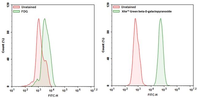
Xite™ Green beta-D-galactopyranoside is a fluorogenic substrate for beta-galactosidase (β-gal). Compared to the existing beta-galactosidase substrates (e.g., the commonly used FDG), it has much better cell permeability. Xite™ Green beta-D-galactopyranoside readily enters cells where it gets cleaved by β-gal, producing Xite™ Green, a strongly fluorescent product. The strongly fluorescent Xite™ Green is well retained in cells, making it easy to be detected with a flow cytometer and fluorescence microscope. Xite™ Green beta-D-galactopyranoside provides a simple and sensitive tool to detect beta-galactosidase activity. Xite™ Green beta-D-galactopyranoside might be used as a simple tool for measuring cellular senescence in cells since β-gal has been identified as a reliable marker for cellular senescence.
Platform
Flow cytometer
| Excitation | 488 nm laser |
| Emission | 530/30 nm filter |
| Instrument specification(s) | FITC channel |
Fluorescence microscope
| Excitation | FITC filter set |
| Emission | FITC filter set |
| Recommended plate | Black wall/clear bottom |
Example protocol
AT A GLANCE
Protocol summary
- Treat samples as desired
- Prepare and add Xite™ Green beta-D-galactopyranoside working solution to samples
- Incubate samples at 37 °C for 15 to 45 minutes
- Monitor the fluorescence intensity using flow cytometer with 530/30 nm filter (FITC channel) or using fluorescence microscopy with FITC filter set
PREPARATION OF STOCK SOLUTIONS
Unless otherwise noted, all unused stock solutions should be divided into single-use aliquots and stored at -20 °C after preparation. Avoid repeated freeze-thaw cycles.
Xite™ Green beta-D-galactopyranoside stock solution
Add appropriate amount of DMSO into Xite™ Green beta-D-galactopyranoside to make 2-5 mM Xite™ Green beta-D-galactopyranoside stock solution. Note: Store the unused Xite™ Green beta-D-galactopyranoside stock solution at -20 °C in single use aliquots.PREPARATION OF WORKING SOLUTION
Xite™ Green beta-D-galactopyranoside working solution
Prepare 1-20 µM of Xite™ Green beta-D-galactopyranoside working solution in buffer of your choice. Note: Xite™ Green beta-D-galactopyranoside working solution should be used promptly. Note: The concentration of the Xite™ Green beta-D-galactopyranoside should be optimized for different cell types and conditions.SAMPLE EXPERIMENTAL PROTOCOL
The following protocol can be used as a guideline and should be optimized according to the needs.
- Treat your samples as desired.
- Remove the treatment and wash the cells with buffer of your choice such as DPBS. Note: For selectively tracking β-Gal in live cells, cells can be treated with Bafilomycin A1 for blocking endogenous β-Gal. Optimum concentration of Bafilomycin A1 may vary on type of cells.
- Add Xite™ Green beta-D-galactopyranoside working solution for 15-45 minutes and incubate the samples at 37 °C incubator. Note: Optimal time for incubation needs to be determined experimentally.
- Remove the working solution and wash cells with buffer of your choice.
- Resuspend the cells in buffer of your choice and monitor the fluorescence intensity with flow cytometer using 530/30 nm filter (FITC channel) or fluorescence microscope with FITC filter set.
Calculators
Common stock solution preparation
Table 1. Volume of DMSO needed to reconstitute specific mass of Xite™ Green beta-D-galactopyranoside to given concentration. Note that volume is only for preparing stock solution. Refer to sample experimental protocol for appropriate experimental/physiological buffers.
| 0.1 mg | 0.5 mg | 1 mg | 5 mg | 10 mg | |
| 1 mM | 202.224 µL | 1.011 mL | 2.022 mL | 10.111 mL | 20.222 mL |
| 5 mM | 40.445 µL | 202.224 µL | 404.449 µL | 2.022 mL | 4.044 mL |
| 10 mM | 20.222 µL | 101.112 µL | 202.224 µL | 1.011 mL | 2.022 mL |
Molarity calculator
Enter any two values (mass, volume, concentration) to calculate the third.
| Mass (Calculate) | Molecular weight | Volume (Calculate) | Concentration (Calculate) | Moles | ||||
| / | = | x | = |
Images

Figure 1. Expression of β-gal was measured with Xite™ Green beta-D-galactopyranoside. 9L-LacZ cells (cells that overexpressed β-gal) were incubated with Xite™ Green beta-D-galactopyranoside or FDG for 30 mins at 37 °C. The signal was acquired with FITC channel using a NovoCyte Flow Cytometer (ACEA Biosciences).
References
View all 18 references: Citation Explorer
Novel fluorescent probe for rapid and ratiometric detection of β-galactosidase and live cell imaging.
Authors: Chen, Xiangzhu and Zhang, Xueyan and Ma, Xiaodong and Zhang, Yuanyuan and Gao, Gui and Liu, Jingjing and Hou, Shicong
Journal: Talanta (2019): 308-313
Authors: Chen, Xiangzhu and Zhang, Xueyan and Ma, Xiaodong and Zhang, Yuanyuan and Gao, Gui and Liu, Jingjing and Hou, Shicong
Journal: Talanta (2019): 308-313
Targeting senescence improves angiogenic potential of adipose-derived mesenchymal stem cells in patients with preeclampsia.
Authors: Suvakov, Sonja and Cubro, Hajrunisa and White, Wendy M and Butler Tobah, Yvonne S and Weissgerber, Tracey L and Jordan, Kyra L and Zhu, Xiang Y and Woollard, John R and Chebib, Fouad T and Milic, Natasa M and Grande, Joseph P and Xu, Ming and Tchkonia, Tamara and Kirkland, James L and Lerman, Lilach O and Garovic, Vesna D
Journal: Biology of sex differences (2019): 49
SA-β-Galactosidase-Based Screening Assay for the Identification of Senotherapeutic Drugs.
Authors: Fuhrmann-Stroissnigg, Heike and Santiago, Fernando E and Grassi, Diego and Ling, YuanYuan and Niedernhofer, Laura J and Robbins, Paul D
Journal: Journal of visualized experiments : JoVE (2019)
Authors: Fuhrmann-Stroissnigg, Heike and Santiago, Fernando E and Grassi, Diego and Ling, YuanYuan and Niedernhofer, Laura J and Robbins, Paul D
Journal: Journal of visualized experiments : JoVE (2019)
Cellular and cytoskeletal alterations of scleral fibroblasts in response to glucocorticoid steroids.
Authors: Bogarin, Thania and Saraswathy, Sindhu and Akiyama, Goichi and Xie, Xiaobin and Weinreb, Robert N and Zheng, Jie and Huang, Alex S
Journal: Experimental eye research (2019): 107774
Authors: Bogarin, Thania and Saraswathy, Sindhu and Akiyama, Goichi and Xie, Xiaobin and Weinreb, Robert N and Zheng, Jie and Huang, Alex S
Journal: Experimental eye research (2019): 107774
Tumor cell escape from therapy-induced senescence.
Authors: Saleh, Tareq and Tyutyunyk-Massey, Liliya and Murray, Graeme F and Alotaibi, Moureq R and Kawale, Ajinkya S and Elsayed, Zeinab and Henderson, Scott C and Yakovlev, Vasily and Elmore, Lynne W and Toor, Amir and Harada, Hisashi and Reed, Jason and Landry, Joseph W and Gewirtz, David A
Journal: Biochemical pharmacology (2019): 202-212
Fluorescent probes for selective protein labeling in lysosomes: a case of α-galactosidase A.
Authors: Bohl, Cornelius and Pomorski, Adam and Seemann, Susanne and Knospe, Anne-Marie and Zheng, Chaonan and Krężel, Artur and Rolfs, Arndt and Lukas, Jan
Journal: FASEB journal : official publication of the Federation of American Societies for Experimental Biology (2017): 5258-5267
Authors: Bohl, Cornelius and Pomorski, Adam and Seemann, Susanne and Knospe, Anne-Marie and Zheng, Chaonan and Krężel, Artur and Rolfs, Arndt and Lukas, Jan
Journal: FASEB journal : official publication of the Federation of American Societies for Experimental Biology (2017): 5258-5267
Identification of a β-galactosidase transgene that provides a live-cell marker of transcriptional activity in growing oocytes and embryos.
Authors: Edwards, Nicole and Farookhi, Riaz and Clarke, Hugh J
Journal: Molecular human reproduction (2015): 583-93
Authors: Edwards, Nicole and Farookhi, Riaz and Clarke, Hugh J
Journal: Molecular human reproduction (2015): 583-93
Characterization of functional capacity of adult ventricular myocytes in long-term culture.
Authors: Liu, Shi J
Journal: International journal of cardiology (2013): 1923-36
Authors: Liu, Shi J
Journal: International journal of cardiology (2013): 1923-36
Abrupt and dynamic changes in gene expression revealed by live cell arrays.
Authors: Walling, Maureen A and Shi, Hua and Shepard, Jason R E
Journal: Analytical chemistry (2012): 2737-44
Authors: Walling, Maureen A and Shi, Hua and Shepard, Jason R E
Journal: Analytical chemistry (2012): 2737-44
Oxidative stress and cell senescence combine to cause maximal renal tubular epithelial cell dysfunction and loss in an in vitro model of kidney disease.
Authors: Small, David M and Bennett, Nigel C and Roy, Sandrine and Gabrielli, Brian G and Johnson, David W and Gobe, Glenda C
Journal: Nephron. Experimental nephrology (2012): 123-30
Authors: Small, David M and Bennett, Nigel C and Roy, Sandrine and Gabrielli, Brian G and Johnson, David W and Gobe, Glenda C
Journal: Nephron. Experimental nephrology (2012): 123-30