logo
AAT Bioquest

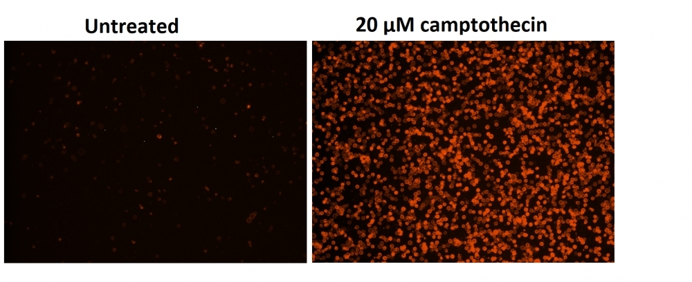
Cell Meter™ Phosphatidylserine Apoptosis Assay Kit *Orange Fluorescence Optimized for Microplate Readers*

This particular kit is designed to monitor cell apoptosis through measuring the translocation of phosphatidylserine (PS). In apoptosis, PS is transferred to the outer leaflet of the plasma membrane. The appearance of phosphatidylserine on the cell surface is a universal indicator of the initial/intermediate stages of cell apoptosis and can be detected before morphological changes can be observed. This kit uses our proprietary orange fluorescent Apopxin™ PS sensor that specifically binds PS with affinity much higher than Annexin V (Kd < 10 nM). The PS sensor used in this kit has orange fluorescence upon binding to membrane PS. The stain has the spectral properties almost identical to those of Cy3® or Alexa Fluor® 555, making it convenient to be used for the common fluorescence instruments equipped with the light sources and filters for Cy3® or Alexa Fluor® 555 (Cy3® or Alexa Fluor® 555 are the trademarks of GE Healthcare and Invitrogen respectively). Due to its highly enhanced affinity to PS, this kit is more robust than the other commercial Annexin V-based apoptosis kits that are only used with either microscope or flow cytometry platform. This kit can be also used with a fluorescence microplate reader besides the microscope and flow cytometry platforms.

Example protocol

AT A GLANCE

Protocol summary

  1. Prepare cells with test compounds (100 µL/well/96-well plate or 25 µL/well/384-well plate)
  2. Add equal volume of Apopxin™ Orange working solution
  3. Incubate at room temperature for 1 hour
  4. Monitor fluorescence intensity (bottom read mode) at Ex/Em = 540/590 nm (Cutoff = 570 nm) or fluorescence microscope with Cy3 filter

Important notes
Warm Assay Buffer (Component B) at room temperature before starting the experiment.

PREPARATION OF WORKING SOLUTION

Add 10 μL of 100X Apopxin™ Orange (Component A) into 1 mL of Assay Buffer (Component B) and mix well to make Apopxin™ Orange working solution. Note: 100 μL of Apopxin™ Orange working solution is enough for one well. Prepare fresh before use.

For guidelines on cell sample preparation, please visit
https://www.aatbio.com/resources/guides/cell-sample-preparation.html

SAMPLE EXPERIMENTAL PROTOCOL

  1. Treat cells with test compounds by adding 10 µL/well (96-well plate) or 2.5 µL/well (384-well plate) of 10X test compound stock solution into PBS or the desired buffer. For blank wells (medium without the cells), add the same amount of compound buffer.

  2. Incubate the cell plate in a 5% CO2, 37°C incubator for a desired period of time (4 - 6 hours for Jurkat cells treated with camptothecin) to induce apoptosis.

  3. Add 100 µL/well (96-well plate) or 25 µL/well (384-well plate) of Apopxin™ Orange working solution into each well.

  4. Incubate the cell plate at room temperature for at least 1 hour, protected from light.

  5. Centrifuge cell plate (especially for the non-adherent cells) at 800 rpm for 2 minutes (brake off).

  6. Monitor the fluorescence intensity with a a fluorescence microplate reader (bottom read mode) at Ex/Em = 540/590 nm (Cutoff = 570 nm) or image cells using fluorescence microscope with Cy3® filter.

Citations

View all 7 citations: Citation Explorer
Receptor tyrosine kinase-like orphan receptor 1 inhibitor strictinin exhibits anti-cancer properties against highly aggressive androgen-independent prostate cancer
Authors: Sivaganesh, Vignesh and Peethambaran, Bela
Journal: Exploration of Targeted Anti-tumor Therapy (2023): 1188--1209
STE20-Type Kinases MST3 and MST4 Act Non-Redundantly to Promote the Progression of Hepatocellular Carcinoma
Authors: Caputo, Mara and Xia, Ying and Anand, Sumit Kumar and Cansby, Emmelie and Andersson, Emma and Marschall, Hanns-Ulrich and K{\"o}nigsrainer, Alfred and Peter, Andreas and Mahlapuu, Margit
Journal: (2023)
Integrin $\beta$3 inhibits hypoxia-induced apoptosis in cardiomyocytes
Authors: Su, Yifan and Tian, Hua and Wei, Lijiang and Fu, Guohui and Sun, Ting
Journal: Acta Biochimica et Biophysica Sinica (2018): 658--665
Tumor-selective mitochondrial network collapse induced by atmospheric gas plasma-activated medium.
Authors: Saito, Kosuke and Asai, Tomohiko and Fujiwara, Kyoko and Sahara, Junki and Koguchi, Haruhisa and Fukuda, Noboru and Suzuki-Karasaki, Miki and Soma, Masayoshi and Suzuki-Karasaki, Yoshihiro
Journal: Oncotarget (2016)

References

View all 92 references: Citation Explorer
Trivalent methylated arsenical-induced phosphatidylserine exposure and apoptosis in platelets may lead to increased thrombus formation
Authors: Bae ON, Lim KM, Noh JY, Chung SM, Kim SH, Chung JH.
Journal: Toxicol Appl Pharmacol (2009): 144
Mobilization of lysosomal calcium regulates the externalization of phosphatidylserine during apoptosis
Authors: Mirnikjoo B, Balasubramanian K, Schroit AJ.
Journal: J Biol Chem (2009): 6918
Evaluation of cell surface expression of phosphatidylserine in ovarian carcinoma effusions using the annexin-V/7-AAD assay: clinical relevance and comparison with other apoptosis parameters
Authors: Dong HP, Holth A, Kleinberg L, Ruud MG, Elstr and MB, Trope CG, Davidson B, Risberg B.
Journal: Am J Clin Pathol (2009): 756
Detection of apoptosis based on the interaction between annexin V and phosphatidylserine
Authors: Liu T, Zhu W, Yang X, Chen L, Yang R, Hua Z, Li G.
Journal: Anal Chem (2009): 2410
Peptidic targeting of phosphatidylserine for the MRI detection of apoptosis in atherosclerotic plaques
Authors: Burtea C, Laurent S, Lancelot E, Ballet S, Murariu O, Rousseaux O, Port M, V and er Elst L, Corot C, Muller RN.
Journal: Mol Pharm (2009): 1903
Page updated on October 24, 2024

Ordering information

Price
Unit size
Catalog Number22794
Quantity
Add to cart

Additional ordering information

Telephone1-800-990-8053
Fax1-800-609-2943
Emailsales@aatbio.com
InternationalSee distributors
Bulk requestInquire
Custom sizeInquire
Technical SupportContact us
Purchase orderSend to sales@aatbio.com
ShippingStandard overnight for United States, inquire for international
Request quotation

Storage, safety and handling

H-phraseH303, H313, H333
Hazard symbolXN
Intended useResearch Use Only (RUO)
R-phraseR20, R21, R22
UNSPSC12352200

Platform

Fluorescence microplate reader

Excitation540 nm
Emission590 nm
Cutoff570 nm
Recommended plateBlack wall, clear bottom
Instrument specification(s)Bottom read mode

Components

Fluorescence images of Jurkat cells in a Costar black wall/clear bottom 96-well plate stained with the Cell Meter&trade; Phosphatidylserine Apoptosis Assay Kit. Jurkat cells were treated without (Left) or with 20 &micro;M camptothecin (Right) for 5 hours. The fluorescence intensity was measured using a fluorescence microscope with&nbsp;Cy3&reg; channel.