logo
AAT Bioquest

Phalloidin-iFluor® 555 Conjugate

This orange fluorescent phalloidin conjugate (equivalent to Alexa Fluor® 555-labeled phalloidin) selectively binds to F-actins. Used at nanomolar concentrations, phalloidin derivatives are convenient probes for labeling, identifying and quantitating F-actins in formaldehyde-fixed and permeabilized tissue sections, cell cultures or cell-free experiments. Phalloidin binds to actin filaments much more tightly than to actin monomers, leading to a decrease in the rate constant for the dissociation of actin subunits from filament ends, essentially stabilizing actin filaments through the prevention of filament depolymerization. Moreover, phalloidin is found to inhibit the ATP hydrolysis activity of F-actin. Phalloidin functions differently at various concentrations in cells. When introduced into the cytoplasm at low concentrations, phalloidin recruits the less polymerized forms of cytoplasmic actin as well as filamin into stable "islands" of aggregated actin polymers, yet it does not interfere with stress fibers, i.e. thick bundles of microfilaments. The property of phalloidin is a useful tool for investigating the distribution of F-actin in cells by labeling phalloidin with fluorescent analogs and using them to stain actin filaments for light microscopy. Fluorescent derivatives of phalloidin have turned out to be enormously useful in localizing actin filaments in living or fixed cells as well as for visualizing individual actin filaments in vitro. Fluorescent phalloidin derivatives have been used as an important tool in the study of actin networks at high resolution. AAT Bioquest offers a variety of fluorescent phalloidin derivatives with different colors for multicolor imaging applications.
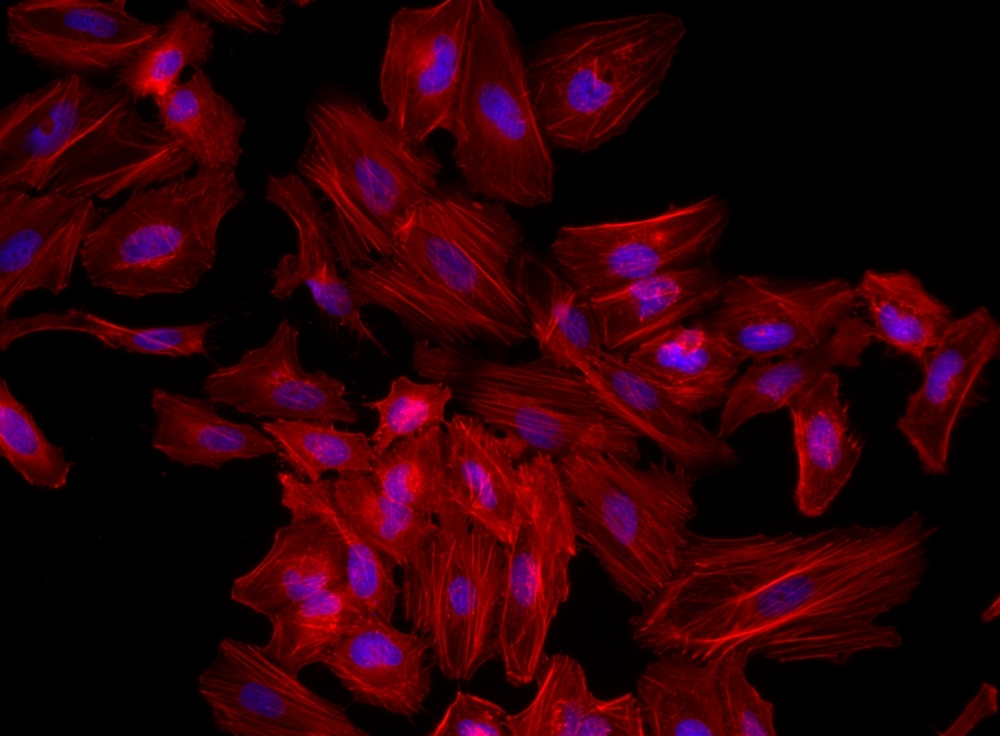
Fluorescence image of HeLa cells fixed with 4% formaldehyde then stained with Phalloidin-iFluor® 555 Conjugate (Cat#23119, Red) and Nuclear Blue™ DCS1 (Cat#17548, Blue), respectively.  
Confocal microscopy images of HEp-2 cells incubated with concentrated culture supernatants of Ssp variants (25 μg/mL) for 5 h. Ssp was visualised with anti-Ssp polyclonal antibody followed by Alexa Fluor Plus 647 conjugated secondary antibody (yellow), actin cytoskeleton was stained with iFluor 555 phalloidin (magenta) and nucleus stained with DAPI (cyan). Ssp variants include wildtype (WT), deletions of Loop 2 (ΔL2), deletion of active site protrusions E2 and E3 (ΔE2, ΔE3, ΔE2/ΔE3), and site-directed mutants of the active site Ser (S314A) and RGD motif (RAE). pBAD denotes vector only control. Images are representative of cells observed from at least three independent experiments. Scale bar represents 50 μm. Source: <b>Crystal structure of a subtilisin-like autotransporter passenger domain reveals insights into its cytotoxic function</b>. by Hor, <em>et. al.</em>., <em>Nature Communications</em>, March 2023.
Axl and integrin αvβ5 expression in AKH tumor cells and glioma cell lines. Cells were cultured on glass slides, fixed with formalin, and then post-treated with Triton-X100. iFluor-555 phalloidin (red) and DAPI (blue) were used to visualize the cells and their nuclei. Bound Axl- and integrin αvβ5-specific monoclonal antibodies were detected by using a secondary anti-mouse antibody-Alexa 488 conjugate (green). Scale = 200 µm. Source: <b>Isolation of Cells from Glioblastoma Multiforme Grade 4 Tumors for Infection with Zika Virus prME and ME Pseudotyped HIV-1</b> by Pöhlking <em>et. al.</em>, <em>Int. J. Mol. Sci.</em> Feb. 2023.
Effects of LPS on filamentous actin arrangement and apoptotic cell death. EA.hy926 cells were incubated with LPS as per the indicated concentrations for 48 h. The treated cell on cell culture slide chamber was stained using fluorescent-tagged phalloidin (Phalloidin-iFluor 555) to visualize the actin filament. Cell images were captured under a fluorescent microscope at 200× magnitude. Treated cells were stained using Annexin V and Dead Cell Reagent (7-AAD) and analyzed using the Muse™ Cell Analyzer. Source: <b>Proteomic Profiling of Early Secreted Proteins in Response to Lipopolysaccharide-Induced Vascular Endothelial Cell EA.hy926 Injury</b> by Songjang, W.; Paiyabhroma, N.; Jumroon, N.; Jiraviriyakul, A.; Nernpermpisooth, N.; Seenak, P.; Kumphune, S.; Thaisakun, S.; Phaonakrop, N.; Roytrakul, S.; <em>et. al. Biomedicines</em>. Nov. 2023
Ordering information
Price
Unit size
Catalog Number23119
Quantity
Add to cart
Additional ordering information
Telephone1-800-990-8053
Fax1-800-609-2943
Emailsales@aatbio.com
InternationalSee distributors
Bulk requestInquire
Custom sizeInquire
ShippingStandard overnight for United States, inquire for international
Request quotation
Physical properties
Molecular weight~1300
SolventDMSO
Spectral properties
Correction Factor (260 nm)0.23
Correction Factor (280 nm)0.14
Extinction coefficient (cm -1 M -1)1000001
Excitation (nm)557
Emission (nm)570
Quantum yield0.641
Storage, safety and handling
Certificate of OriginDownload PDF
H-phraseH301, H311, H331
Hazard symbolT
Intended useResearch Use Only (RUO)
R-phraseR23, R24, R25
StorageFreeze (< -15 °C); Minimize light exposure
UNSPSC12352200
Example protocol

AT A GLANCE

Protocol Summary
  1. Prepare samples in microplate wells
  2. Remove liquid from samples in the plate
  3. Add Phalloidin-iFluor™ 555 Conjugate solution (100 μL/well)
  4. Stain the cells at room temperature for 20 to 90 minutes
  5. Wash the cells
  6. Examine the specimen under microscope with TRITC filter 
Important      Warm the vial to room temperature and centrifuge briefly before opening.

Storage and Handling Conditions
The solution should be stable for at least 6 months if store at -20 °C. Protect the fluorescent conjugates from light, and avoid freeze/thaw cycles.
Note     Phalloidin is toxic, although the amount of toxin present in a vial could be lethal only to a mosquito (LD50 of phalloidin = 2 mg/kg), it should be handled with care.

PREPARATION OF WORKING SOLUTION

Phalloidin-iFluor™ 555 Conjugate working solution
Add 1 µL of Phalloidin-iFluor™ 555 Conjugate solution to 1 mL of PBS with 1% BSA.
Note     The stock solution of phalloidin conjugate should be aliquoted and stored at -20 °C. protected from light.
Note     Different cell types might be stained differently. The concentration of phalloidin conjugate working solution should be prepared accordingly.

SAMPLE EXPERIMENTAL PROTOCOL

Stain the cells
  1. Perform formaldehyde fixation. Incubate cells with 3.0–4.0 % formaldehyde in PBS at room temperature for 10–30 minutes.
    Note     Avoid any methanol containing fixatives since methanol can disrupt actin during the fixation process. The preferred fixative is methanol-free formaldehyde.
  2. Rinse the fixed cells 2–3 times in PBS.
  3. Optional: Add 0.1% Triton X-100 in PBS into fixed cells for 3 to 5 minutes to increase permeability. Rinse the cells 2–3 times in PBS.
  4. Add 100 μL/well (96-well plate) of Phalloidin-iFluor™ 555 Conjugate working solution into the fixed cells, and stain the cells at room temperature for 20 to 90 minutes.
  5. Rinse cells gently with PBS 2 to 3 times to remove excess phalloidin conjugate before plating, sealing and imaging under microscope with TRITC filter set. 
Spectrum
Product family
NameExcitation (nm)Emission (nm)Extinction coefficient (cm -1 M -1)Quantum yieldCorrection Factor (260 nm)Correction Factor (280 nm)
Phalloidin-iFluor® 350 Conjugate3454502000010.9510.830.23
Phalloidin-iFluor® 405 Conjugate4034273700010.9110.480.77
Phalloidin-iFluor® 488 Conjugate4915167500010.910.210.11
Phalloidin-iFluor® 514 Conjugate5115277500010.8310.2650.116
Phalloidin-iFluor® 532 Conjugate5375609000010.6810.260.16
Phalloidin-iFluor® 594 Conjugate58760320000010.5310.050.04
Phalloidin-iFluor® 633 Conjugate64065425000010.2910.0620.044
Phalloidin-iFluor® 647 Conjugate65667025000010.2510.030.03
Phalloidin-iFluor® 680 Conjugate68470122000010.2310.0970.094
Phalloidin-iFluor® 700 Conjugate69071322000010.2310.090.04
Phalloidin-iFluor® 750 Conjugate75777927500010.1210.0440.039
Phalloidin-iFluor® 790 Conjugate78781225000010.1310.10.09
iFluor® 555-streptavidin conjugate55757010000010.6410.230.14
Show More (4)
Citations
View all 118 citations: Citation Explorer
Patterning in stratified epithelia depends on cell--cell adhesion
Authors: Mai, Yosuke and Kobayashi, Yasuaki and Kitahata, Hiroyuki and Seo, Takashi and Nohara, Takuma and Itamoto, Sota and Mai, Shoko and Kumamoto, Junichi and Nagayama, Masaharu and Nishie, Wataru and others,
Journal: Life Science Alliance (2024)
Treponema pallidum protein Tp0136 promotes angiogenesis to facilitate the dissemination of Treponema pallidum
Authors: Li, Wei and Luo, Xi and Zheng, Xin-Qi and Li, Qiu-Ling and Li, Ze and Meng, Qing-Qi and Zeng, Yan-Li and Lin, Yu and Yang, Tian-Ci
Journal: Emerging Microbes \& Infections (2024): 2382236
Multidimensionally Nano-topologized Polycaprolactone Fibrous Membrane Anchored with Bimetallic Peroxide Nanodots for Microenvironment-Switched Treatment on Infected Diabetic Wounds
Authors: Qi, Lin and Huang, Yong and Liu, Zheng and Liu, Jiangshan and Wang, Jing and Xu, Huilun and Yang, Hao and Liu, Limin and Feng, Ganjun and Zhang, Shuyu and others,
Journal: Advanced Fiber Materials (2024): 1--21
Acod1-mediated inhibition of aerobic glycolysis suppresses osteoclast differentiation and attenuates bone erosion in arthritis
Authors: Kachler, Katerina and Andreev, Darja and Thapa, Shreeya and Royzman, Dmytro and Gie{\ss}l, Andreas and Karuppusamy, Shobika and Perez, Mireia Llerins and Liu, Mengdan and Hofmann, J{\"o}rg and Gessner, Arne and others,
Journal: Annals of the Rheumatic Diseases (2024)
Identifying disulfidptosis subtypes in hepatocellular carcinoma through machine learning and preliminary exploration of its connection with immunotherapy
Authors: Chen, Guanjun and Zhang, Ganghua and Zhu, Yuxing and Wu, Anshan and Fang, Jianing and Yin, Zhijing and Chen, Haotian and Cao, Ke
Journal: Cancer Cell International (2024): 194
References
View all 127 references: Citation Explorer
Improved penile histology by phalloidin stain: circular and longitudinal cavernous smooth muscles, dual-endothelium arteries, and erectile dysfunction-associated changes
Authors: Lin G, Qiu X, F and el TM, Albersen M, Wang Z, Lue TF, Lin CS.
Journal: Urology (2011): 970 e1
Phalloidin perturbs the interaction of human non-muscle myosin isoforms 2A and 2C1 with F-actin
Authors: Diensthuber RP, Muller M, Heissler SM, Taft MH, Chizhov I, Manstein DJ.
Journal: FEBS Lett (2011): 767
Labeling cytoskeletal F-actin with rhodamine phalloidin or fluorescein phalloidin for imaging
Authors: Chazotte B., undefined
Journal: Cold Spring Harb Protoc (2010): pdb prot4947
pH-(low)-insertion-peptide (pHLIP) translocation of membrane impermeable phalloidin toxin inhibits cancer cell proliferation
Authors: An M, Wijesinghe D, Andreev OA, Reshetnyak YK, Engelman DM.
Journal: Proc Natl Acad Sci U S A (2010): 20246
Protective effect of bile acid derivatives in phalloidin-induced rat liver toxicity
Authors: Herraez E, Macias RI, Vazquez-Tato J, Hierro C, Monte MJ, Marin JJ.
Journal: Toxicol Appl Pharmacol (2009): 21