logo
AAT Bioquest

Phalloidin-iFluor® 594 Conjugate

This red fluorescent phalloidin conjugate (equivalent to Alexa Fluor® 594-labeled phalloidin) selectively binds to F-actins. Used at nanomolar concentrations, phalloidin derivatives are convenient probes for labeling, identifying and quantitating F-actins in formaldehyde-fixed and permeabilized tissue sections, cell cultures or cell-free experiments. Phalloidin binds to actin filaments much more tightly than to actin monomers, leading to a decrease in the rate constant for the dissociation of actin subunits from filament ends, essentially stabilizing actin filaments through the prevention of filament depolymerization. Moreover, phalloidin is found to inhibit the ATP hydrolysis activity of F-actin. Phalloidin functions differently at various concentrations in cells. When introduced into the cytoplasm at low concentrations, phalloidin recruits the less polymerized forms of cytoplasmic actin as well as filamin into stable "islands" of aggregated actin polymers, yet it does not interfere with stress fibers, i.e. thick bundles of microfilaments. The property of phalloidin is a useful tool for investigating the distribution of F-actin in cells by labeling phalloidin with fluorescent analogs and using them to stain actin filaments for light microscopy. Fluorescent derivatives of phalloidin have turned out to be enormously useful in localizing actin filaments in living or fixed cells as well as for visualizing individual actin filaments in vitro. Fluorescent phalloidin derivatives have been used as an important tool in the study of actin networks at high resolution. AAT Bioquest offers a variety of fluorescent phalloidin derivatives with different colors for multicolor imaging applications.
Example protocol

AT A GLANCE

Protocol Summary
  1. Prepare samples in microplate wells
  2. Remove liquid from samples in the plate
  3. Add Phalloidin-iFluor™ 594 Conjugate solution (100 μL/well)
  4. Stain the cells at room temperature for 20 to 90 minutes
  5. Wash the cells
  6. Examine the specimen under microscope with TRITC or Texas Red filter 
Important      Warm the vial to room temperature and centrifuge briefly before opening.

Storage and Handling Conditions
The solution should be stable for at least 6 months if store at -20 °C. Protect the fluorescent conjugates from light, and avoid freeze/thaw cycles.
Note     Phalloidin is toxic, although the amount of toxin present in a vial could be lethal only to a mosquito (LD50 of phalloidin = 2 mg/kg), it should be handled with care.

PREPARATION OF WORKING SOLUTION

Phalloidin-iFluor™ 594 Conjugate working solution
Add 1 µL of Phalloidin-iFluor™ 594 Conjugate solution to 1 mL of PBS with 1% BSA.
Note     The stock solution of phalloidin conjugate should be aliquoted and stored at -20 °C. protected from light.
Note     Different cell types might be stained differently. The concentration of phalloidin conjugate working solution should be prepared accordingly.

SAMPLE EXPERIMENTAL PROTOCOL

Stain the cells
  1. Perform formaldehyde fixation. Incubate cells with 3.0–4.0 % formaldehyde in PBS at room temperature for 10–30 minutes.
    Note     Avoid any methanol containing fixatives since methanol can disrupt actin during the fixation process. The preferred fixative is methanol-free formaldehyde.
  2. Rinse the fixed cells 2–3 times in PBS.
  3. Optional: Add 0.1% Triton X-100 in PBS into fixed cells for 3 to 5 minutes to increase permeability. Rinse the cells 2–3 times in PBS.
  4. Add 100 μL/well (96-well plate) of Phalloidin-iFluor™ 594 Conjugate working solution into the fixed cells, and stain the cells at room temperature for 20 to 90 minutes.
  5. Rinse cells gently with PBS 2 to 3 times to remove excess phalloidin conjugate before plating, sealing and imaging under microscope with TRITC or Texas Red filter set. 

Spectrum

Product family

NameExcitation (nm)Emission (nm)Extinction coefficient (cm -1 M -1)Quantum yieldCorrection Factor (260 nm)Correction Factor (280 nm)
Phalloidin-iFluor® 350 Conjugate3454502000010.9510.830.23
Phalloidin-iFluor® 405 Conjugate4034273700010.9110.480.77
Phalloidin-iFluor® 488 Conjugate4915167500010.910.210.11
Phalloidin-iFluor® 514 Conjugate5115277500010.8310.2650.116
Phalloidin-iFluor® 532 Conjugate5375609000010.6810.260.16
Phalloidin-iFluor® 555 Conjugate55757010000010.6410.230.14
Phalloidin-iFluor® 633 Conjugate64065425000010.2910.0620.044
Phalloidin-iFluor® 647 Conjugate65667025000010.2510.030.03
Phalloidin-iFluor® 680 Conjugate68470122000010.2310.0970.094
Phalloidin-iFluor® 700 Conjugate69071322000010.2310.090.04
Phalloidin-iFluor® 750 Conjugate75777927500010.1210.0440.039
Phalloidin-iFluor® 790 Conjugate78781225000010.1310.10.09
iFluor® 594-streptavidin conjugate58760320000010.5310.050.04
Show More (4)

Citations

View all 146 citations: Citation Explorer
The SRC family kinase inhibitor NXP900 demonstrates potent anti-tumor activity in squamous cell carcinomas
Authors: Dash, Sweta and Hanson, Sabrina and King, Ben and Nyswaner, Katherine and Foss, Kelcie and Tesi, Noelle and Harvey, Mungo JB and Navarro-Marchal, Sa{\'u}l A and Woods, Allison and Poradosu, Enrique and others,
Journal: Journal of Biological Chemistry (2024): 107615
Hypoxia-Inducible Factor 1$\alpha$ Affects Yak Oocyte Maturation and Early Embryonic Development by Regulating Autophagy
Authors: Ma, Xin and Wang, Meng and Wang, Jinglei and Han, Xiaohong and Yang, Xiaoqing and Zhang, Hui and Zhong, Donglan and Qiu, Shantong and Yu, Sijiu and Wang, Libin and others,
Journal: Antioxidants (2024): 840
Optimizing alkaline hydrothermal treatment for biomimetic smart metallic orthopedic and dental implants
Authors: Hadady, Hanieh and Alam, Arefin and Khurana, Indu and Mutreja, Isha and Kumar, Dhiraj and Shankar, Mamilla Ravi and Dua, Rupak
Journal: Journal of Materials Science: Materials in Medicine (2024): 31
Downregulation of the metalloproteinases ADAM10 or ADAM17 promotes osteoclast differentiation
Authors: Babendreyer, Aaron and Kieselhorst, Julia and Rinkens, Cindy and Lyashenko, Anastasia M and D{\"u}sterh{\"o}ft, Stefan and Jahr, Holger and Craveiro, Rogerio B and Wolf, Michael and Ludwig, Andreas
Journal: Cell Communication and Signaling (2024): 1--15

References

View all 127 references: Citation Explorer
Improved penile histology by phalloidin stain: circular and longitudinal cavernous smooth muscles, dual-endothelium arteries, and erectile dysfunction-associated changes
Authors: Lin G, Qiu X, F and el TM, Albersen M, Wang Z, Lue TF, Lin CS.
Journal: Urology (2011): 970 e1
Phalloidin perturbs the interaction of human non-muscle myosin isoforms 2A and 2C1 with F-actin
Authors: Diensthuber RP, Muller M, Heissler SM, Taft MH, Chizhov I, Manstein DJ.
Journal: FEBS Lett (2011): 767
pH-(low)-insertion-peptide (pHLIP) translocation of membrane impermeable phalloidin toxin inhibits cancer cell proliferation
Authors: An M, Wijesinghe D, Andreev OA, Reshetnyak YK, Engelman DM.
Journal: Proc Natl Acad Sci U S A (2010): 20246
Labeling cytoskeletal F-actin with rhodamine phalloidin or fluorescein phalloidin for imaging
Authors: Chazotte B., undefined
Journal: Cold Spring Harb Protoc (2010): pdb prot4947
Processing of the phalloidin proprotein by prolyl oligopeptidase from the mushroom Conocybe albipes
Authors: Luo H, Hallen-Adams HE, Walton JD.
Journal: J Biol Chem (2009): 18070
Page updated on September 8, 2024

Ordering information

Price
Unit size
Catalog Number23122
Quantity
Add to cart

Additional ordering information

Telephone1-800-990-8053
Fax1-800-609-2943
Emailsales@aatbio.com
InternationalSee distributors
Bulk requestInquire
Custom sizeInquire
Technical SupportContact us
Purchase orderSend to sales@aatbio.com
ShippingStandard overnight for United States, inquire for international
Request quotation

Physical properties

Molecular weight

~1600

Solvent

DMSO

Spectral properties

Absorbance (nm)

587

Correction Factor (260 nm)

0.05

Correction Factor (280 nm)

0.04

Extinction coefficient (cm -1 M -1)

2000001

Excitation (nm)

587

Emission (nm)

603

Quantum yield

0.531

Storage, safety and handling

Certificate of OriginDownload PDF
H-phraseH301, H311, H331
Hazard symbolT
Intended useResearch Use Only (RUO)
R-phraseR23, R24, R25

Storage

Freeze (< -15 °C); Minimize light exposure
UNSPSC12352200
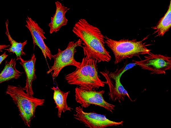
Fluorescence image of HeLa cells fixed with 4% formaldehyde then stained with Cell Navigator® F-Actin Labeling Kit *Red Fluorescence* in a Costar black 96-well plate. Cells were labeled with&nbsp;Phalloidin-iFluor® 594&nbsp; (Cat#23122, Red) and nuclei stain DAPI (Cat#17507, Blue), respectively. Cell endoplasmic reticulum (ER) was stained with ER Green&trade; (Cat#22635, Green) before fixation.
Distribution of MCs in the membranous cochlea. The MCs were scored in each cochlear preparation, and the results are presented as a total number of MCs (non-degranulated and degranulated) per area ((a) n = 7; (b) n = 9). (a) Violin plot demonstrating MCs distribution in the explant areas containing either spiral limbus with OC or the lateral wall; (b) Violin plot demonstrating MCs distribution in the apical, medial, and basal parts of the cochlea; the marker shows the mean. (c–f) Representative micrographs showing the cochlear MCs in the medial part of the cochlea. The explants were stained with phalloidin-iFluor 594, avidin–Alexa Fluor ™ 488, and DAPI. The arrows point to MCs. (f) contains a digital enlargement of the MC image form (e). The data were derived from two independent experiments, and the differences were calculated for means. “ns” indicates not significant (p > 0.05); * p < 0.05; *** p < 0.001. ((a) Kruskal–Wallis test; (b) Mann–Whitney test). Source: <b>Degranulation of Murine Resident Cochlear Mast Cells: A Possible Factor Contributing to Cisplatin-Induced Ototoxicity and Neurotoxicity</b> by Karayay <em>et. al.</em>, <em>Int. J. Mol. Sci.</em> Feb. 2023
Cromolyn used at high concentrations decreases the numbers of IHCs and OHCs. (a) The intact hair cells were scored along the length of 100 µm of the cochlear explant (spiral limbus containing OC), and the percentages of intact IHCs (red circles) and OHCs (green squares) were determined and plotted on the y-axis. The control explants (n = 4) were cultured for 24 h in a tissue culture medium. The treatment groups (n = 4 for each treatment) were cultured for 24 h with cromolyn at the following concentrations: 5 µM, 10 µM, 25 µM, 50 µM, 100 µM, and 200 µM. (b,c) Representative micrograph showing intact (b) and damaged (c) hair cells. Arrows point out intact HCs (white) and damaged HCs (green). Scale bar represents 10 µM. (d–i) Representative micrograph showing the HCs after 24 h of exposure to 5 µM, 10 µM, 25 µM, 50 µM, 100 µM, and 200 µM cromolyn. The cochlea explants were stained with phalloidin-iFluor 594. The scale bar represents 10 µm. Four independent experiments were performed; the data are reported as mean ± SEM. * p < 0.05; ** p < 0.01 (two-way ANOVA with Dunnett multiple comparison test). Source: <b>Degranulation of Murine Resident Cochlear Mast Cells: A Possible Factor Contributing to Cisplatin-Induced Ototoxicity and Neurotoxicity</b> by Karayay <em>et.al.</em>, <em>Int. J. Mol. Sci.</em> Feb. 2023