XFD680 NHS Ester *Same Structure to Alexa Fluor™ 680 NHS Ester*
Product key features
- Ex/Em: 681/704 nm
- Extinction coefficient: 184,000 cm-1M-1
- Reactive Group: NHS ester
- Easy Conjugation: Efficiently labels primary amines on proteins, ligands, and amine-modified oligonucleotides
- Bright & Stable: Delivers intense fluorescence with resilience to photobleaching and pH variations from 4 to 10
- Hydrophilic: Minimizes aggregation, enhancing signal clarity for advanced imaging and live-cell studies
Product description
XFD680, manufactured by AAT Bioquest, is a bright near-infrared fluorescent dye structurally equivalent to Alexa Fluor™ 680 (ThermoFisher). It exhibits a high fluorescence quantum yield, excellent photostability, and superior aqueous solubility, ensuring consistent and reliable performance in various applications. Its pH-independent fluorescence across a broad range (pH 4–11) allows it to maintain stability under diverse experimental conditions. The dye also enables high molar ratio protein conjugation with minimal self-quenching, producing brighter conjugates and enhancing detection sensitivity. Its long-wavelength emission minimizes interference from autofluorescent background signals, enabling accurate detection in complex biological systems.
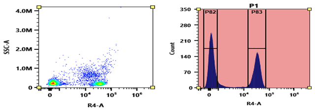
XFD680 is optimized for red laser excitation and is compatible with flow cytometers equipped with spectral detection systems. It delivers robust and uniform labeling, with high signal intensity and reproducibility, making it ideal for fluorescence imaging, flow cytometry, and other analytical techniques. XFD680 is also widely utilized in advanced applications such as multiplexed western blot detection and stochastic optical reconstruction microscopy (STORM), where its superior photophysical properties enhance resolution and sensitivity.
The N-hydroxysuccinimidyl (NHS) ester of XFD680 is a widely used reagent for the conjugation of this dye to proteins or antibodies. NHS esters react selectively and efficiently with primary amines (such as the side chains of lysine residues or aminosilane-coated surfaces) at pH 7-9, forming stable covalent amide bonds. This property makes XFD680 NHS ester an excellent choice for labeling proteins, amine-modified oligonucleotides, and other amine-containing molecules.
Example protocol
PREPARATION OF STOCK SOLUTIONS
Unless otherwise noted, all unused stock solutions should be divided into single-use aliquots and stored at -20 °C after preparation. Avoid repeated freeze-thaw cycles
Prepare a 1 mL protein labeling stock solution by mixing 100 µL of reaction buffer (such as 1 M sodium carbonate solution or 1 M phosphate buffer, pH ~9.0) with 900 µL of the target protein solution (e.g., an antibody with a protein concentration of at least 2 mg/mL, if possible).
Note: The pH of the protein solution (Solution A) should be 8.5 ± 0.5. If the pH of the protein solution is lower than 8.0, adjust it to within the 8.0-9.0 range using either 1 M sodium bicarbonate solution or 1 M phosphate buffer at pH 9.0.
Note: The protein should be dissolved in 1X phosphate-buffered saline (PBS), pH 7.2-7.4. If the protein is dissolved in Tris or glycine buffer, dialyze it against 1X PBS, pH 7.2-7.4, to remove any free amines or ammonium salts (such as ammonium sulfate and ammonium acetate) commonly used in protein precipitation.
Note: Antibodies that are impure or stabilized with bovine serum albumin (BSA) or gelatin may not label effectively. Additionally, sodium azide or thimerosal can interfere with the conjugation reaction. To achieve optimal labeling results, these preservatives should be removed through dialysis or spin column techniques.
Note: For optimal labeling efficiency, it is recommended to maintain a final protein concentration between 2-10 mg/mL. Protein concentrations below 2 mg/mL can significantly reduce conjugation efficiency.
To prepare a 10 mM stock solution of XFD680 NHS ester, add anhydrous DMSO directly to the vial of XFD680 NHS ester. Mix well by pipetting or vortexing.
Note: Prepare the dye stock solution (Solution B) before starting the conjugation, and use it promptly. Extended storage of the dye stock solution may reduce the dye activity. Solution B can be stored in the freezer for up to two weeks, provided it is protected from light and moisture. Avoid freeze-thaw cycles.
SAMPLE EXPERIMENTAL PROTOCOL
This protocol is designed for labeling Goat anti-mouse IgG with XFD680 NHS ester. Additional optimization may be required to adapt the protocol to your specific proteins.
Note: Each protein requires a distinct dye/protein ratio, which varies depending on the characteristics of the dye. Over-labeling a protein can negatively impact its binding affinity, whereas using a low dye-to-protein ratio in protein conjugates can result in reduced sensitivity.
Use a 10:1 molar ratio of Solution B (dye) to Solution A (protein) as the starting point: Add 5 µL of the dye stock solution (Solution B, assuming the dye stock solution is 10 mM) to the vial containing the protein solution (95 µL of Solution A) with effective shaking. The concentration of the protein is ~0.05 mM, assuming the protein concentration is 10 mg/mL and the molecular weight of the protein is ~200KD.
Note: We recommend using a 10:1 molar ratio of Solution B (dye)/Solution A (protein). If it is too low or too high, determine the optimal dye/protein ratio at 5:1, 15:1, and 20:1, respectively.
Continue to rotate or shake the reaction mixture at room temperature for 30-60 minutes.
The following protocol demonstrates the purification of a dye-protein conjugate using a Sephadex G-25 column.
Prepare the Sephadex G-25 column according to the manufacturer's instructions.
Carefully load the reaction mixture (from the "Run Conjugation Reaction" step) to the top of the Sephadex G-25 column.
Add PBS (pH 7.2-7.4) as soon as the sample runs just below the top resin surface.
Add more PBS (pH 7.2-7.4) to the desired sample to complete the column purification. Combine the fractions that contain the desired dye-protein conjugate.
Note: For immediate use, the dye-protein conjugate must be diluted with staining buffer, and aliquoted for multiple uses.
Note: For longer-term storage, the dye-protein conjugate solution needs to be concentrated or freeze-dried.
The Degree of Substitution (DOS) is a critical factor in characterizing dye-labeled proteins. Proteins with a lower DOS generally exhibit weaker fluorescence, while those with a higher DOS (e.g., DOS > 6) may also show reduced fluorescence. The optimal DOS for most antibodies typically ranges between 2 and 10, depending on the specific properties of both the dye and the protein. For effective labeling, it is recommended to achieve a DOS of 6-8 moles of XFD680 NHS ester per mole of antibody. The following steps outline the process for determining the DOS of XFD680 NHS ester-labeled proteins.
For accurate measurement of the absorption spectrum of a dye-protein conjugate, maintain the sample concentration between 1-10 µM, adjusting as needed based on the dye's extinction coefficient.
For most spectrophotometers, the sample (from the column fractions) needs to be diluted with de-ionized water so that the O.D. values are in the range of 0.1 to 0.9. The O.D. (absorbance) at 280 nm is the maximum absorption of protein, while 681 nm is the maximum absorption of XFD680 NHS ester. To obtain accurate DOS, ensure the conjugate is free of the non-conjugated dye.
You can calculate DOS using our tool by following this link:
Spectrum

Product family
| Name | Excitation (nm) | Emission (nm) | Extinction coefficient (cm -1 M -1) | Correction Factor (260 nm) | Correction Factor (280 nm) |
| XFD350 NHS Ester *Same Structure to Alexa Fluor™ 350 NHS Ester* | 343 | 441 | 19000 | 0.25 | 0.19 |
| XFD405 NHS Ester [equivalent to Alexa Fluor™ 405 NHS Ester] | 401 | 421 | 35,000 | 0.23 | 0.70 |
| XFD430 NHS ester | 432 | 540 | 15,000 | - | 0.28 |
| XFD488 NHS Ester *Same Structure to Alexa Fluor™ 488 NHS Ester* | 499 | 520 | 71000 | 0.30 | 0.11 |
| XFD514 NHS Ester *Same Structure to Alexa Fluor™ 514 NHS Ester* | 518 | 543 | 80000 | 0.31 | 0.18 |
| XFD532 NHS Ester *Same Structure to Alexa Fluor™ 532 NHS Ester* | 534 | 553 | 81000 | 0.24 | 0.09 |
| XFD546 NHS Ester *Same Structure to Alexa Fluor™ 546 NHS Ester* | 561 | 572 | 112000 | 0.21 | 0.12 |
| XFD555 NHS Ester *Same Structure to Alexa Fluor™ 555 NHS Ester* | 553 | 568 | 150000 | 0.08 | 0.08 |
| XFD568 NHS Ester *Same Structure to Alexa Fluor™ 568 NHS Ester* | 579 | 603 | 91300 | 0.45 | 0.46 |
Show More (18) | |||||
Citations
Authors: Patil, M. K., Kotresh, M. G., Inamdar, S. R.
Journal: Spectrochim Acta A Mol Biomol Spectrosc (2019): 142-152
Authors: Ostad, S. N., Babaei, S., Bayat, A. A., Mahmoudian, J.
Journal: Monoclon Antib Immunodiagn Immunother (2019): 25-29
Authors: Rai, S., Bhardwaj, U., Misra, A., Singh, S., Gupta, R.
Journal: Int J Lab Hematol (2018): e52-e54
Authors: Velazquez-Lopez, I., Leon-Cruz, E., Pardo, J. P., Sosa-Peinado, A., Gonzalez-Andrade, M.
Journal: Anal Biochem (2017): 13-22
Authors: Cui, J. J., Zhu, X. L., Ji, C. F., Jing, X. H., Bai, W. Z.
Journal: Zhen Ci Yan Jiu (2011): 262-7
References
Authors: Velazquez-Lopez, I.; Leon-Cruz, E.; Pardo, J. P.; Sosa-Peinado, A.; Gonzalez-Andrade, M.
Journal: Anal Biochem (2017): 13-22
Authors: Winne, J. M.; Irani, N. G.; Van den Begin, J.; Madder, A.
Journal: Methods Mol Biol (2017): 21-Sep
Authors: Maroteaux, M.; Liu, S. J.
Journal: eNeuro (2016)
Authors: Grate, J. W.; Mo, K. F.; Shin, Y.; Vasdekis, A.; Warner, M. G.; Kelly, R. T.; Orr, G.; Hu, D.; Dehoff, K. J.; Brockman, F. J.; Wilkins, M. J.
Journal: Bioconjug Chem (2015): 593-601
Authors: Fenton, K. E.; Martirosyan, N. L.; Abdelwahab, M. G.; Coons, S. W.; Preul, M. C.; Scheck, A. C.
Journal: Neurosurg Focus (2014): E12