Cell Cycle Assays
Flow cytometric measurements of cellular DNA content are commonly used to assess DNA ploidy level and to measure the percentages of a cell population in the different phases of the cell cycle. In a given population, the distribution of cells within the major phases of the cell cycle - G0/G1, S, and G2/M - are based on the differences in their DNA content. By using stoichiometric, DNA binding dyes that exhibit emission signals proportional to the amount of DNA present in each cell, FACS analysis of stained populations can be used to generate frequency histograms that estimate the percentages of cells in each cell cycle phase. Cell cycle analysis can be used to investigate tumor behavior and detect variations in growth patterns, monitor for apoptosis, and evaluate the effect of various pharmacologic agents on cell cycle progression. While analysis is typically performed on cells that have been permeabilized and/or fixed using membrane-impermeant DNA dyes, AAT Bioquest offers live cell cycle reagents ideal for cell sorting applications, immunophenotyping, and multiplex analysis with other live-cell studies.
Live Cell Cycle Assays for Flow Cytometry
The Cell Meter™ Fluorimetric Live Cell Cycle Assay Kits provide a simple and sensitive method for monitoring cell cycle progression in live, permeabilized, and fixed cells. Each Cell Meter™ Fluorimetric Live Cell Cycle Assay Kit contains a patented membrane-permeant and non-fluorescent, DNA-selective dye. Upon binding to DNA, these dyes emit fluorescence signals proportional to the DNA content present in each cell. Flow cytometric analysis of the stained population can then be used to generate a frequency histogram (Figure 1) that provides information regarding the relative percentage of cells in the major phases of the cell cycle.

DNA profile in growing and nocodazole treated Jurkat cells. Jurkat cells were treated without (A) or with 100 ng/mL Nocodazole (B) in a 37 °C, 5% CO2 incubator for 24 hours, then incubated with Nuclear Green™ LCS1 for 30 minutes. The fluorescence intensity of Nuclear Green™ LCS1 was measured with an ACEA NovoCyte flow cytometer in the FITC channel. In growing Jurkat cells (A), nuclear staining with Nuclear Green™ LCS1 shows G1, S, and G2 phases. In Nocodazole treated G2 arrested cells (B), the frequency of G2 cells increased dramatically, while G1 and S phase-frequency decreased significantly.
The DNA dyes provided in the Cell Meter™ Fluorimetric Live Cell Cycle Assay Kits are designed to have absorption spectra that match the primary wavelengths of standard laser lines. Nuclear Violet™ - the DNA dye component in Kit 22845 - is well-suited for the 405 nm (violet) laser line but can also be excited by the 355 nm (UV) laser. Nuclear Green™ LCS1 and Nuclear Red™ CCS2 - the DNA dye components in Kit 22841 and Kit 22860, respectively - are well-suited for the 488 nm (blue) laser line. Nuclear Red™ CCS2 can also be excited using the 532 nm (green) laser line. In addition to convenience, the non-cytotoxicity of Nuclear DNA dyes enables multiplexing with additional live cell applications, including immunophenotyping, analysis of GFP cells, FACS, CFSE cell tracing, and apoptotic sub-G1 population analysis. The Nuclear DNA dyes are also available as stand-alone reagents in 0.5 mL unit sizes, and in a variety of other fluorescence emissions, including Nuclear Yellow (Ex/Em = 368/501 nm, 17539), Nuclear Orange™ LCS1 (Ex/Em = 512/554 nm, 17541), Nuclear Red™ LCS1 (Ex/Em = 622/645 nm, 17542), and Nuclear Red™ LCS2 (Ex/Em = 651/681 nm, 17545).

Absorbance spectra of Nuclear Violet™, Nuclear Green™ LCS1, and Nuclear Red™ CCS2 DNA dyes provided in the Cell Meter™ Fluorimetric Live Cell Cycle Assay Kits. Nuclear Violet™ is well excited by the 405 nm laser (violet) and the 355 nm laser (black). Nuclear Green™ LCS1 is well excited by the 488 nm laser (blue). Nuclear Red™ CCS2 is well excited by the 488 nm laser (blue) and the 532 nm laser (green).
Cell Meter™ Fluorimetric Live Cell Cycle Assay Kits selection guide
| Cell Meter Fluorimetric Live Cell Cycle Assay Kit *Optimized for 405 nm Violet Laser Excitation* | Cell Meter Fluorimetric Live Cell Cycle Assay Kit *Green Fluorescence Optimized for Flow Cytometry* | Cell Mete Fluorimetric Live Cell Cycle Assay Kit *Red Fluorescence Optimized for Flow Cytometry* | |
| Dye Component | Nuclear Violet™ (view spectrum) | Nuclear Green™ LCS1 (view spectrum) | Nuclear Red™ CCS2 (view spectrum) |
| Ex/Em (nm) | 401/460 | 503/527 | 542/580 |
| Laser (nm) | UV or Violet (355 or 405 nm) | Blue (488 nm) | Blue or Green (488 or 532 nm) |
| Cytotoxicity | Non-cytotoxic | Non-cytotoxic | Non-cytotoxic |
| Multiplex | Yes | Yes | Yes |
| Unit Size | 100 Tests | 100 Tests | 100 Tests |
| Cat No. | 22845 | 22841 | 22860 |
Fixed Cell Cycle Assays for Flow Cytometry
For fixed cell cycle analysis, AAT Bioquest offers a Cell Meter™ Fluorimetric Fixed Cell Cycle Assay Kit (22842) optimized for flow cytometry, as well as classic cell cycle dyes such as DAPI (17511), propidium iodide (17515), and 7-AAD (17501). The dye component in Kit 22842 - Nuclear Red™ CCS1 - is a membrane-impermeant, blue laser-excited DNA dye. Upon binding to DNA in fixed cells, Nuclear Red™ CCS1 exhibits significant fluorescence enhancement, with signal intensities proportional to the DNA content present in each cell. Because Nuclear Red™ CCS1 also binds to RNA, RNase A treatment (kit component B) is required for DNA content analysis. Nuclear Red™ CCS1 has a peak emission of ~618 nm that can be detected using the same filter for PE-Texas Red, such as a 610/20 bandpass filter.
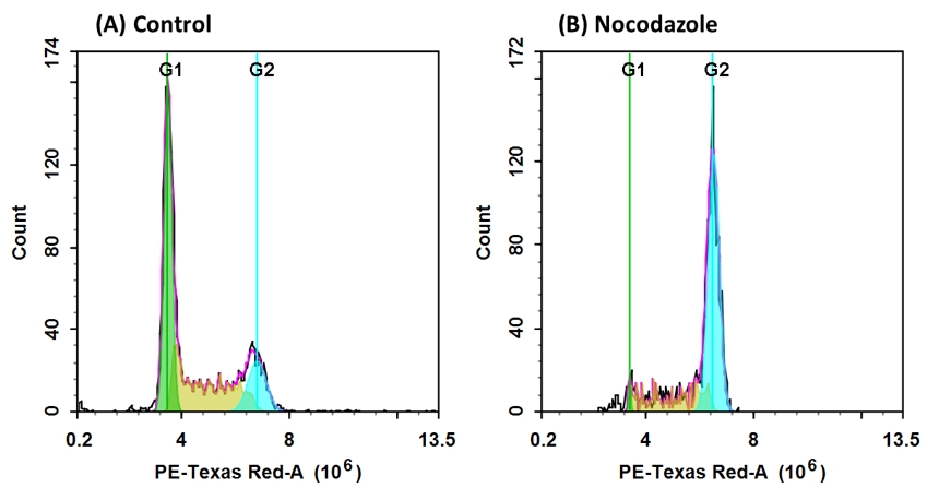
DNA profile in growing and nocodazole treated Jurkat cells. Jurkat cells were treated without (A) or with 100 ng/mL Nocodazole (B) in a 37 °C, 5% CO2 incubator for 24 hours before fixation with 70% ethanol. Cells were then loaded with Nuclear Red™ CCS1 for 30 minutes and treated with RNase A. The fluorescence intensity of Nuclear Red™ CCS1 was measured with an ACEA NovoCyte flow cytometer in the PE-Texas Red channel. In growing Jurkat cells (A), nuclear staining with Nuclear Red™ CCS1 shows G1, S, and G2 phases. In Nocodazole treated G2 arrested cells (B), the frequency of G2 cells increased dramatically, while G1 and S phase-frequency decreased significantly.
Cell Meter™ Fluorimetric Fixed Cell Cycle Assay Kit guide
| Cell Meter Fluorimetric Fixed Cell Cycle Assay Kit *Red Fluorescence Optimized for Flow Cytometry* | |
| Dye Component | Nuclear Red CCS1 (view spectrum) |
| Ex/Em (nm) | 537/618 |
| Laser (nm) | Blue or Green (488 or 532 nm) |
| Multiplex | Yes |
| Unit Size | 100 Tests |
| Cat No. | 22842 |
Hoechst, PI, and Other Classic Cell Cycle Stains
Nucleic acid stains commonly used for cell-cycle analysis, include Hoechst 33258 (17520, 17523, 17525), Hoechst 33342 (17530, 17533, 17535), DAPI (17507, 17510, 17511, 17513), PI (17515, 17516, 17517) and 7-AAD (17501). The Hoechst stains and DAPI are UV laser-excited dyes with emissions around 460 nm, while PI and 7-AAD take advantage of the commonly available 488 nm excitation sources with emissions at 618 nm and 648 nm, respectively. Of these classic dyes, Hoechst 33342 and Hoechst 33258 are suitable for live cell-cycle analysis, although Hoechst 33258 is reportedly less cell-permeant. Cell fixation and permeabilization is, however, a requirement for cell cycle analysis when using DAPI, PI, or 7-AAD. For dyes like PI, which are not DNA specific and will stain other double-stranded nucleic acids such as RNA, treatment with RNase A is necessary for DNA content analysis.
Classic cell cycle dyes selection Guide
| Dye | Hoechst 33258 | Hoechst 33342 | DAPI | PI | 7-AAD |
| Ex/Em (nm) | 352/454 | 352/454 | 359/457 | 537/618 | 549/648 |
| Laser (nm) | UV (355 nm) | UV (355 nm) | UV (355 nm) | Blue or Green (488 or 532 nm) | Blue or Green (488 or 532 nm) |
| Bandpass Filter (nm) | 450/50 | 450/50 | 450/50 | 610/10 | 670/20 |
| Live or Fixed Cells | For live and fixed cells | For live and fixed cells | For fixed cells | For fixed cells | For fixed cells |
| Unit Size (Cat No.) | 100 mg (17520) 1 g (17523) 5 mL (17525) | 100 mg (17530) 1 g (17533) 5 mL (17535) | 2 mL (17507) 10 mg (17510) 25 mg (17513) 100 mg (17511) | 25 mg (17515) 5 g (17516) 1 mL (17517) | 1 mg (17501) |
Product Ordering Information
Table 1. Cell cycle assays and reagents for flow cytometry.
| Product ▲ ▼ | Unit Size ▲ ▼ | Cat No. ▲ ▼ |
| 7-AAD [7-Aminoactinomycin D] *CAS 7240-37-1* | 1 mg | 17501 |
| Cell Navigator® CDy6 Mitosis Imaging Kit | 100 tests | 22640 |
| Cell Meter™ Fluorimetric Fixed Cell Cycle Assay Kit *Optimized for 405 nm Violet Laser Excitation* | 100 tests | 22842 |
| Cell Meter™ Fluorimetric Live Cell Cycle Assay Kit *Green Fluorescence Optimized for Flow Cytometry* | 100 tests | 22841 |
| Cell Meter™ Fluorimetric Live Cell Cycle Assay Kit *Optimized for 405 nm Violet Laser Excitation* | 100 tests | 22845 |
| Cell Meter™ Fluorimetric Live Cell Cycle Assay Kit *Red Fluorescence Optimized for Flow Cytometry* | 100 tests | 22860 |
| DAPI [4,6-Diamidino-2-phenylindole, dihydrochloride] *10 mM solution in water* | 2 mL | 17507 |
| DAPI [4,6-Diamidino-2-phenylindole, dihydrochloride] *CAS 28718-90-3* | 10 mg | 17510 |
| DAPI [4,6-Diamidino-2-phenylindole, dihydrochloride] *CAS 28718-90-3* | 25 mg | 17513 |
| DAPI [4,6-Diamidino-2-phenylindole, dihydrochloride] *CAS 28718-90-3* | 100 mg | 17511 |