Cell Signaling Pathways
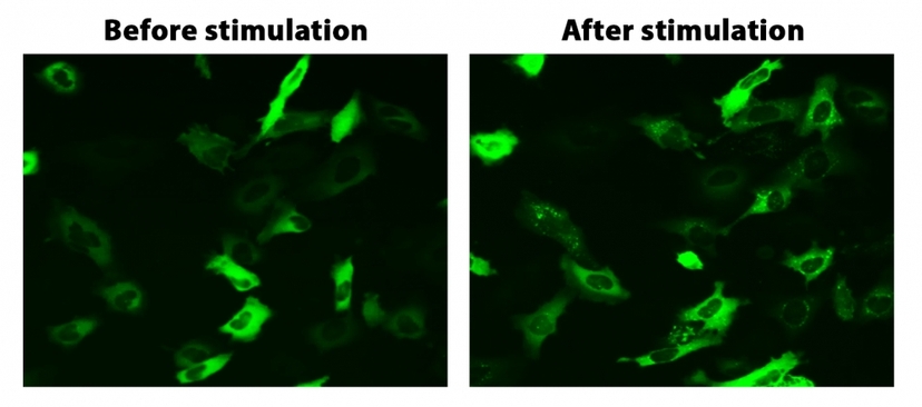
Translocation of beta-arrestin in HeLa cells. HeLa cells were transiently transfected with beta-arrestin-GFP and vasopressin receptor 2 (V2R). HeLa cells were cultured in a 6-well plate and grown to ~60% confluence. Equal amounts of beta-arrestin-GFP (1.5 µg) and V2R plasmids (1.5 µg) were transfected with 9 µL of Transfectamine™ 5000. Cells were transferred to a 96-well plate ~ 30 hours after transfection. Vasopressin (1 µM) was added to the cells ~ 48 hours after transfection to induce beta-arrestin-GFP translocation. Images were taken before and 2 hours after the vasopressin treatment under a fluorescent microscope using the FITC channel.
Cells will receive a signal, or activate, when ligands like cytokines, growth factors, or hormones bind to specific protein receptors on the cell surface. Individual cells are likely to receive many signals simultaneously, and all of this information will become integrated into a unified process. After the first molecule in the pathway is triggered, it activates the next molecule in the path. This process is repeated through a chain of communication until a final cell is activated and exhibits a functional response. This operation, termed signal transduction, occurs through tightly organized networks of protein-protein interactions and signaling complexes controlled by firmly regulated domains.
Because signaling pathways play vital roles in cellular function, differentiation, and coordination in the body, it is no surprise that abnormal activation or dysfunction of these pathways is linked to numerous diseases. Therefore, elucidating cell signal pathways has become paramount in advancing drug discovery and for developing a better understanding of pathophysiologic and pharmacologic mechanisms.
Types of Cell Signaling
Most cell signals are chemical in nature, and can either exert their effects locally or travel distances to reach their targets from their source of origin. Multicellular organisms have a number of cells that backpack chemical signals from one location to the next. Possibly the best-known examples are neurotransmitters, a category of short-range signaling molecules that travel across the spaces between neurons and/or muscle cells to facilitate movement and thought. Hormones, however, travel from the brain all the way to other organs, like the ovary, where it can trigger egg release.
Additionally, some cells may respond to mechanical stimuli. This can easily be seen in how sensory cells in the skin respond to the pressure of touch, or specialized vascular cells can detect changes in blood pressure which is used to maintain a consistent cardiac load. Prokaryotic organisms, too, have sensors that can detect and react to chemical, mechanical, thermal, or even electromagnetic stimuli. As of today, there are five main categories of signal types in multicellular organisms.
Autocrine Signals
Autocrine signals are produced by cells that can bind to a ligand that is released after activation. In autocrine signaling, the signaling and target cell can be of the same type, or similar. This signaling often occurs during early development to ensure cells differentiate correctly and take on proper function. Similar neighboring cells can also be influenced by the released autocrine ligand, which can help direct embryogenesis thus ensuring the correct developmental outcome. Autocrine signaling also regulates pain sensation and inflammatory responses in immune cells. Additionally, if a cell is infected with a virus, the cell can use autocrine signals to undergo programmed cell death, killing the virus in the process.
Juxtacrine Signals
Juxtacrine signaling is based on the interaction of noncleaved ligand precursors and the epidermal growth factor receptor (EGFR). Juxtacrine ligands are anchored to a membrane and target adjacent cells, where signaling requires the juxtaposition of ligands and receptors on the surfaces of the exosome and target cell. There exist three types of juxtacrine interactions, the first two of which occur when a protein on one cell can bind to the receptor of another, or a receptor on one cell can bind to the extracellular matrix. Thirdly, it is also possible for a juxtacrine signal to be transmitted directly from the cytoplasm of one cell directly into the cytoplasm of an adjacent cell.
Juxtacrine signaling plays a key role in the development of the nervous system, from neurogenesis through axonal growth. Such signaling is also used in neurexin-neuroligin interactions to direct synaptogenesis and synaptic remodeling. It also directs some T cell and checkpoint receptors towards antigens to help regulate immune response.
Paracrine Signaling
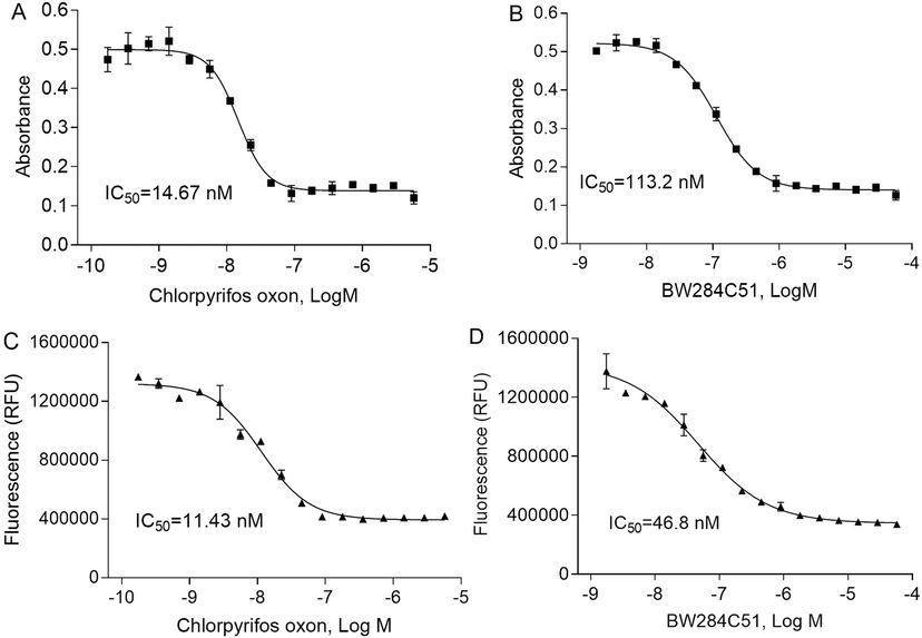
Concentration-response curves of human AChE assays. There were concentration-dependent inhibition curves in 1536-well plates treated with two positive controls, chlorpyrifos oxon and BW284C5. Two methods were used to measure AChE activity including colorimetric (A, B) and fluorescent methods (C, D). Each value represents the mean±SD of three independent experiments. Amplite Colorimetric Acetylcholinesterase Assay kit (Ellman assay) and Amplite Fluorimetric Acetylcholinesterase Assay kit (Green Fluorescence) from AAT Bioquest. Source: Use of high-throughput enzyme-based assay with xenobiotic metabolic capability to evaluate the inhibition of acetylcholinesterase activity by organophosphorous pesticides by Li et.al., Toxicology in Vitro. April 2019.
This reaction helps to maintain a localized response, and one example of paracrine signals are in the synapses between nerve cells that are propagated by fast-moving electrical impulses. First, these impulses travel from the cell body to the tips of their axons, which are long armlike extensions. Next, the signal continues on to a dendrite, a short branched arm, of the next cell through the release of chemical ligands, known as neurotransmitters, by the presynaptic cell. The speed of nerve signaling is facilitated by the small distance between nerve cells, which enables fast response. Other key examples of paracrine signals include responses to allergens, blood clotting, tissue repair, and the formation of scar tissue by keratinocytes and fibroblasts.
Endocrine ligands, or hormones, are molecules that are produced in one part of the body, but frequently affect other regions throughout the entire organism. Hormones travel through the bloodstream, a mechanism that results in a slow response, but with a longer-lasting effect. Traveling through the bloodstream also dilutes the hormones, so they are usually in reduced concentrations when they finally act on target cells. There are multiple examples of endocrine signaling within the body. For example, the pineal gland secretes melatonin which controls the sleep-wake cycle within the brain and also affects the ovaries, blood vessels, and gastrointestinal tract. As well, the pituitary gland releases many reproductive and growth hormones, while the thyroid gland releases many hormones that help control metabolism. Additionally, the hypothalamus releases oxytocin and vasopressin which affects the kidneys and blood vessels; such signals are paramount to controlling water retention and vascular control.
Intracrine signals are those that are produced by a cell and bind to an internal receptor within the signal-producing cell to incorporate the direct action of growth factors. This signaling pattern creates an internal loop that requires the presence of suitable intracellular active receptors. The mechanisms supporting intracrine growth control are less known, and intracrine signaling has been suggested as a potential mechanism for the biological activity of secretory growth factors. Some growth factors may produce dual factor-receptor complexes at the cell surface which are then rapidly internalized and translocated to the nucleus without degradation.
Intracrine signals may also be involved in local, cell- and tissue-specific regulation of sex steroid production and metabolism. Most of these locally produced steroids are primarily generated from circulating, adrenal-derived steroid precursors. Intracrinology recognizes this duality; there is an extensive capacity of virtually all cell types to both generate and metabolize some steroids and thereby tightly regulate signaling in an autonomous fashion.
Table 1. Amplite® Neurotransmitter and Nerve Cell Signaling Assay Kits
| Cat# ▲ ▼ | Product Name ▲ ▼ | Ex ▲ ▼ | Em ▲ ▼ | Unit Size ▲ ▼ |
| 13620 | Amplite® Colorimetric Sphingomyelinase Assay Kit *Blue Color* | 655 | 200 Tests | |
| 13621 | Amplite® Fluorimetric Sphingomyelinase Assay Kit *Red Fluorescence* | 571 | 584 | 200 Tests |
| 13622 | Amplite® Fluorimetric Acidic Sphingomyelinase Assay Kit *Red Fluorescence* | 571 | 584 | 200 Tests |
| 13625 | Amplite® Fluorimetric Sphingomyelin Assay Kit *Red Fluorescence* | 571 | 585 | 100 Tests |
| 11400 | Amplite® Colorimetric Acetylcholinesterase Assay Kit | 410 | 200 Tests | |
| 11401 | Amplite® Fluorimetric Acetylcholinesterase Assay Kit *Green Fluorescence* | 510 | 525 | 200 Tests |
| 11402 | Amplite® Fluorimetric Acetylcholinesterase Assay Kit *Red Fluorescence* | 571 | 585 | 200 Tests |
| 11403 | Amplite® Fluorimetric Acetylcholine Assay Kit *Red Fluorescence* | 571 | 585 | 200 Tests |
Endocrine Signals
Endocrine ligands, or hormones, are molecules that are produced in one part of the body, but frequently affect other regions throughout the entire organism. Hormones travel through the bloodstream, a mechanism that results in a slow response, but with a longer-lasting effect. Traveling through the bloodstream also dilutes the hormones, so they are usually in reduced concentrations when they finally act on target cells. There are multiple examples of endocrine signaling within the body. For example, the pineal gland secretes melatonin which controls the sleep-wake cycle within the brain and also affects the ovaries, blood vessels, and gastrointestinal tract. As well, the pituitary gland releases many reproductive and growth hormones, while the thyroid gland releases many hormones that help control metabolism. Additionally, the hypothalamus releases oxytocin and vasopressin which affects the kidneys and blood vessels; such signals are paramount to controlling water retention and vascular control.
Intracrine signaling
Intracrine signals are those that are produced by a cell and bind to an internal receptor within the signal-producing cell to incorporate the direct action of growth factors. This signaling pattern creates an internal loop that requires the presence of suitable intracellular active receptors. The mechanisms supporting intracrine growth control are less known, and intracrine signaling has been suggested as a potential mechanism for the biological activity of secretory growth factors. Some growth factors may produce dual factor-receptor complexes at the cell surface which are then rapidly internalized and translocated to the nucleus without degradation.
Intracrine signals may also be involved in local, cell- and tissue-specific regulation of sex steroid production and metabolism. Most of these locally produced steroids are primarily generated from circulating, adrenal-derived steroid precursors. Intracrinology recognizes this duality; there is an extensive capacity of virtually all cell types to both generate and metabolize some steroids and thereby tightly regulate signaling in an autonomous fashion.
Signal Transduction
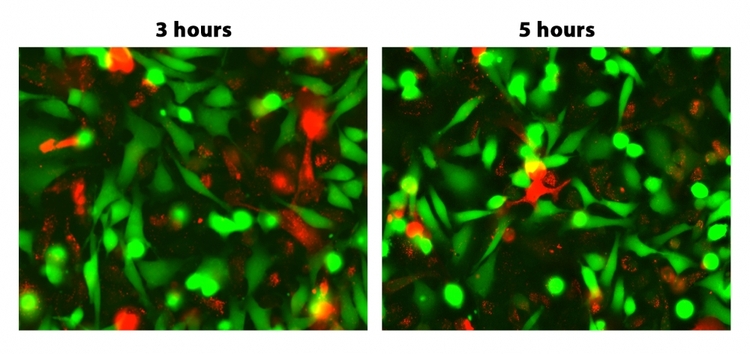
GAP junctions were analyzed by fluorescence microscopy. HeLa cells were stained with Calcein Ultragreen AM and DiD separately as per the protocol. Cells were mixed well with 1:1 ratio and replated with cell culture medium. Images were acquired using a fluorescence microscope with the FITC and Cy5 filter sets. As time progresses, double positive population of Calcein Ultragreen AM and DiD increases.
Signal transduction mechanisms frequently involve second messengers which are small molecules that transmit signals from activated receptors to the cell interior. This interaction can result in changes in the expression of genes and the activity of enzymes. One prominent example is cyclic AMP (cAMP), a second messenger commonly involved in numerous signal transduction cascades. When certain signals reach cAMP molecules, these molecules have the capability to trigger the enzyme protein kinase A (PKA), which then undergoes active phosphorylation on multiple substrates. The signal is amplified with each step, which allows PKA phosphorylation reactions to mediate both short- and long-term cell responses. On a cellular level, signal transduction influences numerous cellular processes including cell growth, division, differentiation, migration, metabolism, immune response, neuronal communication, and death.
In the bigger picture, signal transduction is essential for sensory processes, such as vision and smell, which are used by multicellular organisms to perceive their environment. Even unicellular organisms depend on cell signal transduction to detect chemical cues related to the presence of competitors, predators, toxic environments, and nutrients. In many methods of signal transduction, organisms will induce changes on the target cell via interacting with cell receptors.
Table 2. ELISA For cAMP Assay Kits
| Cat# ▲ ▼ | Product Name ▲ ▼ | Unit Size ▲ ▼ | Ex ▲ ▼ | Em ▲ ▼ |
| 36373 | Screen Quest™ Fluorimetric ELISA cAMP Assay Kit | 1 plate | 571 | 585 |
| 36374 | Screen Quest™ Fluorimetric ELISA cAMP Assay Kit | 10 plates | 571 | 585 |
Cell Receptors
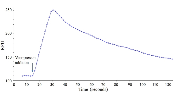
Vasopressin responses in CHO cells as assessed with the Screen Quest™ Live Cell cAMP Assay Service Pack. Vasopressin (50 µL/well) was added using FlexStation 3 to achieve the final concentration of 100 nM.
Although many receptors exist on the exterior of the cell, some are within the cytoplasm, and some even inside the nucleus. Such receptors typically bind to molecules that can pass through the plasma membrane, like nitrous oxide or some steroid hormones. Once a receptor receives a signal it initiates a signal transduction cascade through a conformational change, and this small change normally produces multiple intracellular signals. Though several receptor subtypes have evolved to be directly involved in signal transduction, there also exist receptors that do not have any intrinsic biochemical activity. For example, B cell receptors, T cell receptors, integrins, and interleukin receptors typically play a by-standing role to cooperate with other proteins, instead of directly involving themselves with the signal transduction process.
Cell Signaling and Disease
Cell signaling pathways are involved in the pathophysiology of many diseases, and the nature of these defects and how they are induced varies enormously. Some commonly known diseases and their relation to cell signaling pathways involve two methods of cancer. In these methods, either the growth-inhibiting, tumor suppressing, pathways are inactivated, or the growth-promoting, oncogenic, pathways are activated. Additionally, diabetes is known to arise from multiple defects in insulin signaling pathways involved in blood glucose homeostasis.
Pathogenic organisms and viruses can also interfere with cell signaling pathways, thereby causing adverse function, though this pathophysiology is less clear. Other serious diseases, such as hypertension, heart disease, and many forms of mental illness, seem to arise from subtle changes in signaling pathways. For these reasons, the mechanistic action of many drugs relies on the understanding of cell signaling pathways. Knowledge of disease pathophysiology can therefore be essential to pharmacologic development. Though progress in this area has been enhanced by the completion of the human genome project, an ongoing challenge in cell signaling is to understand the temporal and spatial regulation of signaling events within, and between, cells.
| Application Notes: | FAQs: | |
Techniques for Identifying Signal Pathways
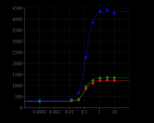
Comparison of fluorescent signal response of endogenous P2Y receptor to ATP in CHO-K1 cells. Calcium flux response was measured with Screen Quest™ Calbryte™ 520 Probenecid-Free and Wash-Free Calcium Assay Kit, FLIPR Calcium 4 Assay Kit , and Fluo-4 Direct Calcium Assay kit. ATP (50 µL/well) was added by FlexStation 3 to achieve the final indicated concentrations.
In experimentation, cell samples are loaded with ratiometric or single-wavelength dyes and then analyzed for analyte binding. Intracellular free calcium concentrations or pH changes can shed light on signal pathways, and can be analyzed by standard laboratory equipment like fluorescence microscopy, flow cytometry, and fluorescence spectroscopy.
Table 3. Cell Meter™ Calcium Assay Kits and Other Calcium Indicators
| Product Name ▲ ▼ | Ex (nm) ▲ ▼ | Em (nm) ▲ ▼ | Unit Size ▲ ▼ | Cat No. ▲ ▼ |
| Cell Meter™ Flow Cytometric Calcium Assay Kit | 493 | 515 | 100 Tests | 36310 |
| Cell Meter™ No Wash and Probenecid-Free Endpoint Calcium Assay Kit *Optimized for microplate reader* | 495 | 525 | 100 Tests | 36312 |
| OG488 BAPTA-1, hexapotassium salt [equivalent to Oregon Green® 488 BAPTA-1, hexapotassium salt] *Cell impermeant* | 493 | 522 | 500 µg | 20506 |
| OG488 BAPTA-1, AM [equivalent to Oregon Green® 488 BAPTA-1, AM] *Cell permeant* | 493 | 522 | 10x50 µg | 20507 |
Immunohistochemistry can likewise be used to locate signaling proteins, the location of which may indicate whether the protein is activated. A variety of antibodies are commercially available for various immunohistochemical techniques. Some antibodies have recognition epitopes that include the phosphate or other activating conformations, other antibodies work against active epitopes to identify signal transduction proteins.
Conversely, it may also be preferred that cells undergo double-labeling with an antibody specific for the target protein and another that recognizes all serine, threonine, or tyrosine phosphorylated amino acids. Another visualization technique involves the construction of fusion proteins, like green fluorescent protein (GFP), caged proteins, or other similar fluorescent or epitope tags. Here, vectors with the tagged-molecule track the expression pathway of analytes by fusing to the target protein and becoming transfected into cells, tissues, or transgenic animals. Yet, other common methods of a visualization assay involve Förster resonance energy transfer (FRET) and fluorescence recovery after photobleaching (FRAP). FRET and FRAP are two imaging techniques that have recently re-emerged as useful methods for advanced visualization of GFP-protein and similar constructs.
Alternative to visualization assays, there exist assays that work by assessing cellular physicochemical response, essentially cell behavior. These assays work by assessing differences in cellular characteristics, including cytoskeletal configuration, cell shape changes, molecular binding, migration, and even differentiation. Many times, assays that assess cell behavior are used to determine whether up- or down-regulating specific signal transduction proteins will impact a change to cellular behavior. In experimentation, transduction proteins are modulated by specific inhibitors to intracellular kinases or cell surface receptors. Among other things, these experiments include assays to assess cell migration and invasion, angiogenesis, reactive oxygen species, protein, and live cell analysis.
Lastly, cell signal pathways may also be assessed by blocking specific kinases or receptors and then assessing activity by using inhibitors. Such inhibitors directly influence and interact with specific molecules that are highly incorporated in various cell signaling pathways, underlining their potential for use as pharmacologic agents. Some inhibitors may simply affect kinases or other reactive enzymes at one concentration, but when the concentration is increased it will affect a wider class of molecules. Some inhibitors can act as competitive inhibitors for ATP binding sites where some inhibition may even be reversible. Many inhibitors are commercially available for both generalized and specific interactions with either various receptors or specific kinases.
| Datasets: | Application Notes: | Assaywise Letters: |
Product Ordering Information
Table 4. Screen Quest™ Calcium Assays for screening GPCR and calcium channel targets.
| Calcium Assay ▲ ▼ | Ex (nm) ▲ ▼ | Em (nm) ▲ ▼ | Cutoff (nm) ▲ ▼ | Unit Size ▲ ▼ | Cat No. ▲ ▼ |
| Screen Quest™ Fluo-4 No Wash Calcium Assay Kit | 490 | 525 | 515 | 10 plates | 36325 |
| Screen Quest™ Fluo-4 No Wash Calcium Assay Kit | 490 | 525 | 515 | 100 plates | 36326 |
| Screen Quest™ Fluo-8 No Wash Calcium Assay Kit | 490 | 525 | 510 | 1 plates | 36314 |
| Screen Quest™ Fluo-8 No Wash Calcium Assay Kit | 490 | 525 | 510 | 10 plates | 36315 |
| Screen Quest™ Fluo-8 No Wash Calcium Assay Kit | 490 | 525 | 510 | 100 plates | 36316 |
| Screen Quest™ Fluo-8 No Wash Calcium Assay Kit | 490 | 525 | 510 | 100 plates | 36316 |
| Screen Quest™ Fluo-8 Medium Removal Calcium Assay Kit *Optimized for Difficult Cell Lines* | 490 | 525 | 510 | 1 plates | 36307 |
| Screen Quest™ Fluo-8 Medium Removal Calcium Assay Kit *Optimized for Difficult Cell Lines* | 490 | 525 | 510 | 10 plates | 36308 |
| Screen Quest™ Fluo-8 Medium Removal Calcium Assay Kit *Optimized for Difficult Cell Lines* | 490 | 525 | 510 | 100 plates | 36309 |
| Screen Quest™ Calbryte-520 Probenecid-Free and Wash-Free Calcium Assay Kit | 490 | 525 | 515 | 1 plates | 36317 |
Table 5. Biochemical assays for monitoring the activation of adenylyl cyclase in G-protein coupled receptor systems.
| Assay ▲ ▼ | Ex/Abs (nm) ▲ ▼ | Em (nm) ▲ ▼ | Cutoff (nm)¹ ▲ ▼ | Microplate Type ▲ ▼ | Unit Size ▲ ▼ | Cat No. ▲ ▼ |
| Screen Quest™ Fluorimetric ELISA cAMP Assay Kit | 540 | 590 | 570 | Solid black plate | 1 plates | 36373 |
| Screen Quest™ Fluorimetric ELISA cAMP Assay Kit | 540 | 590 | 570 | Solid black plate | 10 plates | 36374 |
| Screen Quest™ Colorimetric ELISA cAMP Assay Kit | 405, 650, 740 | - | - | Clear plate | 1 plate | 36370 |
| Screen Quest™ Colorimetric ELISA cAMP Assay Kit | 405, 650, 740 | - | - | Clear plate | 10 plates | 36371 |
| Screen Quest™ FRET No Wash cAMP Assay Kit | - | - | - | Solid black plate, black wall/clear bottom plate | 1 plates | 36379 |
| Screen Quest™ FRET No Wash cAMP Assay Kit | - | - | - | Solid black plate, black wall/clear bottom plate | 10 plates | 36380 |
| Screen Quest™ FRET No Wash cAMP Assay Kit | - | - | - | Solid black plate, black wall/clear bottom plate | 50 plates | 36381 |
Table 6. Probes For Detecting And Monitoring cGAMP Activity
| Cat# ▲ ▼ | Product Name ▲ ▼ | Unit Size ▲ ▼ |
| 20310 | 2',3'-cGAMP | 100 µg |
| 20311 | 2',3'-cGAMP | 1 mg |
| 20313 | 2',3'-cGAMP, sodium salt | 100 µg |
| 20316 | 2',3'-cGAMP-Biotin conjugate | 100 µg |
| 20318 | 2',3'-cGAMP-Cy5 conjugate | 100 µg |
| 20320 | 2’,3’-cGAMP-iFluor 488 conjugate | 100 µg |
| 20322 | 2’,3’-cGAMP-iFluor 647 conjugate | 100 µg |
| 20330 | 2’,3’-cGAMP-DBCO conjugate | 100 µg |
| 20332 | 2’,3’-cGAMP azide | 100 µg |
| 20334 | 2’,3’-cGAMP succinimidyl ester | 100 µg |
Table 7. Related Kinase Products for Protein Kinase Activity
| Cat# ▲ ▼ | Product Name ▲ ▼ | Unit Size ▲ ▼ |
| 11410 | Amplite® Colorimetric Enterokinase Activity Assay Kit | 200 Tests |
| 31001 | Amplite® Universal Fluorimetric Kinase Assay Kit *Red Fluorescence* | 250 Tests |
| 31005 | ReadiUse™ Universal Kinase Reaction Stopping Buffer | 100 mL |
| 8A0459 | Aurora Kinase (Phospho-Thr288) Antibody | - |
| 8A0531 | p70 S6 Kinase (Phospho-Thr229) Antibody | - |
| 8A0532 | p70 S6 Kinase (Phospho-Ser371) Antibody | - |
| 8A0533 | p70 S6 Kinase (Phospho-Thr389) Antibody | - |
| 8A0534 | p70 S6 Kinase (Phospho-Ser418) Antibody | - |
| 8A0761 | Fructose 6 Phosphate Kinase (Phospho-Ser483) Antibody | - |
| 8A0783 | Breast Tumor Kinase (Phospho-Tyr447) Antibody | - |
References
Cell Signaling
Signal Transduction
Signaling Molecules and Cellular Receptors - Forms of Signaling
An intracrine view of sex steroids, immunity, and metabolic regulation
Juxtacrine Signaling
Signaling Molecules and Their Receptors
Signalling Defects and Disease
Approaches To Studying Cellular Signaling: A Primer For Morphologists
Juxtacrine Signalling
Paracrine Signalling
