Protein Assays & Analysis

Three-fold dilution series of BSA standards were separated on a NuPAGE® 4-12% Bis-Tris gel and stained with A) ProLite™ Orange Protein Gel Stain or B) Coomassie brilliant blue (CBB) according to standard protocols. The ProLite™ Orange stained gels were photographed using a SYPRO Orange filter. The CBB-stained gels were photographed using transmitted white light without an optical filter. Lane 1: 15ug, Lane 7: ~20ng, Lane 10: ~0.8 ng BSA.
Protein assays can also be a final detection step in the clinical laboratory as part of disease diagnosis, for example in identifying concentration of hormones, cytokines or enzymes associated with a specific metabolic state or pathological variations. Many assays are available for assessing total protein, specific proteins, or an array of various protein concentrations. Other protein assays might also incorporate mass determination of proteins, the testing of their activity, the determination of the exact amino acid sequence, or for their intrinsic interaction between molecules.
Bradford Assay

BSA dose responses were measured with Amplite® Colorimetric Bradford Protein Quantitation Assay Kit using a clear bottom 96-well plate. A) Detection with A595/A460nm. B) Detect with A595 nm as an alternative option if A460 nm is not available. The Bradford assay tends to lend itself well to adaption.
Table 1. Bradford Protein Assays
| Cat# ▲ ▼ | Product Name ▲ ▼ | Unit Size ▲ ▼ |
| 11118 | Amplite® Colorimetric Bradford Protein Quantitation Assay Kit | 1000 Tests |
| 11119 | Amplite® Colorimetric Bradford Protein Quantitation Assay Kit | 5000 Tests |
The Bradford assay offers fast experimental times that can be performed at room temperature, the assay does not require complex or hazardous chemicals, and results are stable for up to one hour. Importantly, the starting material must have an appropriate number of target residues or the dye will not bind efficiently, resulting in underestimated protein concentration. Though this assay is also incompatible with most detergents like SDS, commonly used to solubilize membrane proteins, Bradford assay-compatible reagents are commercially available. Contaminating detergents can also be removed by gel filtration, dialysis, precipitation with calcium phosphate, or acetone, though these additional steps usually cause sample dilution or loss.
| Tools: | FAQs: |
Lowry Assay
The Lowry assay is a total protein quantitation assay that can detect protein concentration in a range between 10-1000 ug/ml. The Lowry assay works on the principle of copper chelation, where reduced copper is targeted then detected through colorimetry. In a typical experimental setup, first copper and nitrogenous protein interact under alkaline conditions. This interaction, known as the Biuret reaction, reduces copper ions which form complexes with peptide bonds in residues. Next, the complexed residues, specifically tyrosine and tryptophan, react with a Folin-Ciocalteu phenol reagent that produces a blue or green color. The color produced is relatable to the concentration of protein within the sample, and detectable with a spectrophotometer in the range of 650 to 750 nm. Protein concentration can be estimated by creating a standard curve from a known, common protein (e.g., bovine serum albumin (BSA)).
The Lowry assay is highly sensitive and accurate, especially when analyzed at 750 nm since fewer substances absorb light at this wavelength. As the Lowry assay is an endpoint assay, one standard curve can be used for a number of comparable experiments. The Lowry assay, however, is not compatible with reducing agents (e.g., DTT, β-mercaptoethanol) or potassium and magnesium ions. Many compounds commonly used in buffers for protein preparation (e.g., detergents, carbohydrates, glycerol, Tricine, EDTA, Tris) can interfere with protein interactions and form precipitates, so these should be avoided.
| Tools: |
The Bicinchoninic Acid (BCA) Assay
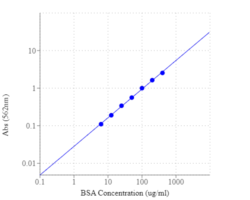
BSA dose responses were measured with Amplite® Colorimetric BCA Protein Quantitation Assay Kit using a clear bottom 96-well plate.
The BCA assay is extremely sensitive, and has even been used to detect total protein concentration as low as 5 ug/mL. This assay type offers low variability and more uniform response than other conventional assays, and can easily be done in a 96-well plate, providing more potential for high throughput applications. The BCA assay is primarily involved with the peptide backbone, and is less affected by differences in amino acid composition. Yet, the BCA assay comes with some limitations. Some reagents, like EDTA, reducing sugars and lipids, or chemicals that interact with copper (e.g., ammonia) can interfere with the reaction. These interferences can be somewhat ameliorated by dialysis, gel filtration or if the protein concentration is high enough, sample dilution. The presence of chemically modified residues in the protein may also interfere with the BCA assay. For example, one experiment showed that protein concentrations in highly methylated samples were consistently overestimated (Brady and Macnaughtan, 2019).
Table 2. Bicinchoninic Acid (BCA) Protein Assays
| Cat# ▲ ▼ | Product Name ▲ ▼ | Unit Size ▲ ▼ |
| 11115 | Amplite® Colorimetric BCA Protein Quantitation Assay Kit | 1000 Tests |
| 11116 | Amplite® Colorimetric BCA Protein Quantitation Assay Kit | 5000 Tests |
UV Spectroscopic Protein Assays
UV spectroscopy is a relatively nonspecific, total protein assessment that can be performed using a spectrophotometer set to an absorbance at 280 nm. This method measures the characteristic absorption of the aromatic residues tyrosine, tryptophan, and phenylalanine. Concentration can be derived from knowing the number or proportion of aromatic side chains present in a sample. For pure protein samples, the exact amino acid sequence must be known and the protein-specific absorption coefficient must be calculated prior to determining protein concentration, using Beer-Lambert law:
A = ε l c
A = Absorbance @ 280nm
ε = molar extinction coefficient
l = path length of the spectrometer
c = molar concentration of protein
| Tools: |
Enzyme-linked immunosorbent assay (ELISA)

Representation of direct and indirect antigen detection using target specific antibodies in ELISA.
In the ELISA reaction, a colorimetric change is reported as an optical density, proportional to the amount of a captured antigen in the sample. ELISA kits are commercially available, inexpensive, and include straightforward protocols. There are four common variations in technique (direct, indirect, sandwich, and competitive ELISAs), making ELISA a dynamic and effective tool in research. Additionally, ELISA systems can be automated and even multiplexed.
Table 3. General ELISA Kits For Product Ordering Information
| Cat# ▲ ▼ | Product Name ▲ ▼ | Ex (nm) ▲ ▼ | Em (nm) ▲ ▼ | Size ▲ ▼ |
| 11540 | Amplite® Fluorimetric Goat Anti-Mouse IgG-HRP Conjugate ELISA Assay Kit *Red Fluorescence* | 571 | 585 | 1000 Tests |
| 11541 | Amplite® Fluorimetric Goat Anti-Rabbit IgG-HRP Conjugate ELISA Assay Kit *Red Fluorescence* | 571 | 585 | 1000 Tests |
| 36370 | Screen Quest™ Colorimetric ELISA cAMP Assay Kit | 650 | 1 plate | |
| 36371 | Screen Quest™ Colorimetric ELISA cAMP Assay Kit | 650 | 10 plates | |
| 36373 | Screen Quest™ Fluorimetric ELISA cAMP Assay Kit | 571 | 585 | 1 plate |
| 36374 | Screen Quest™ Fluorimetric ELISA cAMP Assay Kit | 571 | 585 | 10 plates |
| 36379 | Screen Quest™ TR-FRET No Wash cAMP Assay Kit | 390 | 650 | 1 plate |
| 36380 | Screen Quest™ TR-FRET No Wash cAMP Assay Kit | 390 | 650 | 10 plates |
| 36381 | Screen Quest™ TR-FRET No Wash cAMP Assay Kit | 390 | 650 | 50 plates |
| V101000 | Amplite® Human Apolipoprotein A1 (ApoA1) Kit *Optimized For ELISA Development with ALP* | 96 Tests |
Western Blot

P53 Antibody Western Blot, validation (1:1000 dilution) in T8.
These primary antibodies can either be monoclonal or polyclonal, and if preferred, a secondary antibody can also be included to aid in the detection, sorting or purification of target antigen. Next, the blots can be processed through exposure to an autoradiographic film. Analysis is simple, as positively labeled proteins will appear as dark bands on the film. Each step in Western blot must be optimized, and these steps combined can be a more lengthy process.
Table 4. Enzyme-labeled secondary antibodies for western blotting.
| Cat# ▲ ▼ | Product Name ▲ ▼ | Unit Size ▲ ▼ |
| 11035 | MegaWox™ polyHRP-Goat Anti-Mouse IgG Conjugate | 1 mg |
| 11037 | MegaWox™ polyHRP-Goat Anti-Rabbit IgG Conjugate | 1 mg |
| 16728 | HRP-labeled goat anti-mouse IgG (H+L) | 1 mg |
| 16793 | HRP-labeled goat anti-rabbit IgG (H+L) | 1 mg |
| 50260 | HRP Goat Anti-human IgG (H+L) Antibody | 200 ug |
| 50261 | HRP Goat Anti-human IgG (H+L) Antibody | 1 mg |
| 50262 | HRP Goat Anti-human IgG (H+L) Antibody *Cross Adsorbed* | 200 ug |
| 50263 | HRP Goat Anti-human IgG (H+L) Antibody *Cross Adsorbed* | 1 mg |
Mass Spectrometry
Protein mass spectrometry can be used to characterize, qualitate, and quantify proteins in a single-step experiment. Several mass spectrometry-based workflows have been developed for protein quantitation, including those for labeling metabolites, proteins, chimeric recombinant proteins, peptides, isobaric peptides, and even synthetic peptides. A typical experiment includes a handful of steps, the first of which involves protein labeling. In labeling, the protein sample is labeled with a stable heavy isotope (e.g., 13C or 15N) while the internal standard is labeled with a light isotope (e.g., 12C or 14N). Then, the protein and standard are mixed and the sample undergoes lysis, fractionation, and lastly digestion.
After, the sample undergoes analysis by a mass analyzer. In investigation, each labeled protein forms a peak, and the area under the peak corresponds to their relative abundance of sample protein and the standard. Additionally, multiple internal protein standards can be used in tandem in a single experiment, enabling capabilities for multiplexing. Though mass spectrometry is incredibly accurate, it requires costly instrumentation which has severely limited its utility in research and development.
| FAQs: | Calculators: | Digital Catalogs: |
Fluorescent Assays

BSA dose response was measured on a solid black 96-well plate with Amplite® Fluorimetric Fluorescamine Protein Quantitation Assay Kit.
Some fluorescent assays will link the protein of interest to a fluorescent tag that has a particular cis-acting hydrolase element, known as a protein quantitation reporter. This technique ensures stoichiometric expression of both the protein and the reporter, since protein quantity can be assessed from the intensity of the fluorescent signal. One increasingly common reporter includes green fluorescent protein (GFP), and in research GFP-tagged proteins have proven useful as a means to quantitatively measure the expression of a specific protein. Fluorescent assays are not harmful to living cells, so have the added advantage for use in vitro.
Table 5. Fluorescent Protein Quantitation Assays
| Cat# ▲ ▼ | Product Name ▲ ▼ | Unit Size ▲ ▼ |
| 11100 | Amplite® Fluorimetric Fluorescamine Protein Quantitation Kit *Blue Fluorescence* | 200 Tests |
| 11105 | Amplite® Fluorimetric Protein Quantitation Kit *Orange Fluorescence* | 500 Tests |
| 11109 | Portelite™ Fluorimetric Protein Quantitation Kit *Optimized for CytoCite™ and Qubit™ Fluorometers* | 100 Tests |
| 11111 | Portelite™ Fluorimetric Protein Quantitation Kit *Optimized for CytoCite™ and Qubit™ Fluorometers* | 500 Tests |
Microarrays
Protein microarrays can be triply used for protein quantification, to elucidate protein interaction partners, and to track post translational modifications. Techniques are often described as forward microarrays, where the forward- or normal-phase approach first consists of the immobilization of a capture molecule (e.g., antibodies or aptamers) to a surface. Microarrays can be performed with purified proteins, antibodies, or a cell extract, and experiments can be done either by directly labeled proteins or, in case of an immobilized prey antibody, with a second antibody that recognizes the target protein.
A great advantage of microarray technology is that it offers the possibility for miniaturization and parallelization; basically, that many samples may be quantitated for specific proteins side-by-side in a rapid, low sample volume format. In proteomics, protein and antibody microarrays have been used to assess the up- and downregulation of numerous biomarkers involved in diseases, like cancer. This research may be essential for progressing drug development in that tumors can be more efficiently targeted, so that they can be eliminated quicker and also prevent metastasis.
Nanoparticles and Quantum Dots

Example of quantitation of surface amino groups on magnetic nanoparticle with Beadlite™ Rapid Colorimetric Amino Quantitation Kit.
Notably, quantum dots are a type of nanoparticle made from semiconducting materials. The dots show quantum effects because of their miniscule size, where electrons inside the dot are trapped and can only occupy defined energy levels. Assays that utilize quantum dots have shown outstanding photoluminescent properties, which make them significant antigen-detection systems.
Table 6. Beadlite™ Rapid Colorimetric Amino Quantitation Kit for Nanoparticles
| Cat# ▲ ▼ | Product Name ▲ ▼ | Unit Size ▲ ▼ |
| 5532 | Beadlite™ Rapid Colorimetric Amino Quantitation Kit for Nanoparticles | 2 Tests |
| Application Notes: | FAQs: | Resources: |
Product Ordering Information
Table 7. Fluorescent Protein Stains
| Cat# ▲ ▼ | Product Name ▲ ▼ | Unit Size ▲ ▼ |
| 18000 | ProLite™ Orange Protein Gel Stain *5000X* | 100 µL |
| 18001 | ProLite™ Orange Protein Gel Stain *5000X* | 1 mL |
| 18002 | ProLite™ FAST Blue Protein Gel Stain *200 Gels* | 1 Set |
Table 8. Amplite® Quantitation Kits For Proteins in Solution
| Cat# ▲ ▼ | Product Name ▲ ▼ | Ex ▲ ▼ | Em ▲ ▼ | Unit Size ▲ ▼ |
| 11100 | Amplite® Fluorimetric Fluorescamine Protein Quantitation Kit *Blue Fluorescence* | 380 | 470 | 200 Tests |
| 11105 | Amplite® Fluorimetric Protein Quantitation Kit *Orange Fluorescence* | 485 | 590 | 500 tests |
| 11115 | Amplite® Colorimetric BCA Protein Quantitation Assay Kit | 562 | 1000 tests | |
| 11118 | Amplite® Colorimetric Bradford Protein Quantitation Assay Kit | 595 | 1000 tests |
References
Top 5 Protein Quantification Assays
Protein Quantitation
Antibody Applications
Protein assay
Determination of conjugated protein on nanoparticles by an adaptation of the Coomassie blue dye method
Fluorescent labeling and modification of proteins
The Principles and Applications of Avidin-Based Nanoparticles in Drug Delivery and Diagnosis
Protein Quantitation Using Mass Spectrometry