logo
AAT Bioquest

iFluor® 460 maleimide

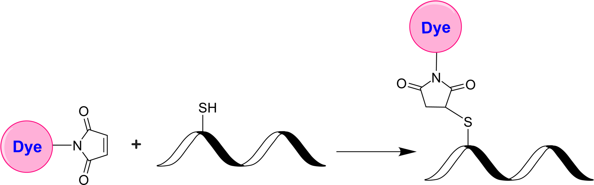
AAT Bioquest's iFluor® dyes are optimized for labeling proteins, particularly antibodies. These dyes are bright, photostable, and have minimal quenching on proteins. Although the 460 nm blue diode laser is being installed in numerous new fluorescence instruments, few dyes can be well excited at 460 nm. iFluor® 460 is optimized to be well excited by the blue diode laser at 460 nm, enabling new biological applications for the new fluorescence instruments equipped with the 460 nm blue diode laser. iFluor® 460 maleimide is stable and shows good reactivity and selectivity with the thiol group.

Example protocol

PREPARATION OF STOCK SOLUTIONS

Unless otherwise noted, all unused stock solutions should be divided into single-use aliquots and stored at -20 °C after preparation. Avoid repeated freeze-thaw cycles

Prepare iFluor® 460 maleimide stock solution
  1. Allow the vial of iFluor® dye maleimide to warm up to room temperature.
  2. Add anhydrous DMSO to the vial to prepare a 10 mM dye stock solution.
  3. Vortex the vial briefly to fully dissolve the dye, and then centrifuge to collect the dye at the bottom of the vial.
  4. Protect all stock solutions from light as much as possible by wrapping containers in aluminum foil.
Prepare antibody or protein solution for labeling
  1. If your protein already contains a thiol group, prepare the protein at 50-100 uM (for example: 5mg/ml BSA is ~75uM) in 50~100 mM MES buffer or buffers of your choice with pH 6.5~7.0.
  2. If labeling with an intact antibody, reduction of disulfide bonds need to be carried out before maleimide reaction. Prepare antibody in 2-10 mg/ml in a suitable buffer with pH 7.0–7.5. A 10-fold molar excess of a reducing agent such as DTT or TCEP is added to the antibody. If DTT is used, it must be removed by dialysis or desalting to a suitable buffer with pH 6.5~7.0 prior to conjugation. If TCEP is used, it is not necessary to remove excess TCEP during conjugation with maleimides, however, removal of TCEP by dialysis or desalting prior to conjugation gives the better labeling efficiency.

    Below is a sample protocol for generating free thiol groups on antibody:
    1. Prepare 2-10mg/ml IgG solution in PBS.
    2. Prepare a fresh solution of 1 M DTT (15.4 mg/100 µL) in distilled water. 
    3. Add 1- 20 µL of DTT stock per ml of IgG solution while mixing. 
    4. Let the solution stand at room temperature for 30 minutes without additional mixing (to minimize the re-oxidation of cysteines to cystines). 
    5. Pass the reduced IgG over a filtration column pre-equilibrated with 50 mM MES buffer (pH=6.5) to remove excess DTT.
    6. Determine the antibody concentrations. This can be done either spectrophotometrically or colorimetrically.
    7. Carry out the conjugation as soon as possible after this step.

    Note: For the best results, IgG solutions should be > 2 mg/mL.

    Note: The reduction can be carried out in almost any buffer from pH 7 to 7.5, e.g., MES, phosphate, or TRIS buffers.

    Note: Steps 5 can be replaced by dialysis.

     
  3. If your protein doesn’t have a free thiol group or disulfide bond to reduce, a thiolation modification need to be carried out before maleimide conjugation (for example:  using 2-Iminothiolane or 2-IT) to introduce sulfhydryl (-SH) groups to the original amino groups on protein.

SAMPLE EXPERIMENTAL PROTOCOL

This labeling protocol was developed for the labeling IgG with iFluor® Dye maleimide. Further optimization may be required for your specific proteins.

Note: Each protein requires a distinct dye/protein ratio, which also depends on the properties of dyes. Over-labeling of a protein could detrimentally affect its binding affinity while the protein conjugates of low dye/protein ratio give reduced sensitivity.

Run Conjugation Reaction
  1. Use a 10~20:1 molar ratio of iFluor® dye maleimide : IgG as the starting point. While stirring or vortexing the protein solution, add a volume of dye stock solution to result in a dye: protein molar ratio of 10-20. For example, for 5mg/ml IgG (~33 uM), you would add dye to a final concentration of 0.33-0.66 mM.

    Note: We recommend using a 10:1 molar ratio of dye to protein.  If the ratio is too low or too high, determine the optimal dye/protein ratio at 5:1, 15:1, and 20:1, respectively.
  2. Continue to rotate or shake the reaction mixture at room temperature for 30-60 minutes.
Purify the Conjugation
The following protocol is an example of dye-protein conjugate purification by using a Sephadex G-25 column.
  1. Purify the conjugate on a gel filtration column, such as a Sephadex G-25 column or equivalent matrix, or by extensive dialysis at 4°C in an appropriate buffer.

Recommended AAT Desalting Columns:

Volume of Reaction Catalog#
0.6-1.0mL

Cat#60504: PD-10 Column

https://www.aatbio.com/products/readiuse-disposable-pd-10-desalting-column?unit=60504
~0.1mL

Cat#60500: Spin Column

https://www.aatbio.com/products/readiuse-bio-gel-p-6-spin-column?unit=60500 

Optional: Characterize the Desired Dye-Protein Conjugate

Determining the Degree of Substitution (DOS) is crucial in characterizing dye-labeled proteins. Lower DOS proteins tend to have weaker fluorescence, but higher DOS proteins may also have reduced fluorescence. For most antibodies, the optimal DOS is between 2 and 10, depending on the dye and protein properties. For effective labeling, the degree of substitution should be controlled to have 5-8 moles of iFluor® 460 maleimide to one mole of antibody. The following steps are used to determine the DOS of iFluor® 460 maleimide-labeled proteins:

  1. Measure absorption— To measure the absorption spectrum of a dye-protein conjugate, the sample concentration should be kept between 1 and 10 µM (For example: IgG conjugate: 10uM is ~1.5mg/ml), depending on the dye's extinction coefficient. 
  2. Read OD (absorbance) at 280 nm and dye maximum absorption (ƛ max = 468 nm for iFluor® 460 dyes). For most spectrophotometers, the sample (from the column fractions) must be diluted with de-ionized water so that the OD values range from 0.1 to 0.9. The O.D. (absorbance) at 280 nm is the maximum absorption of protein, while 468 nm is the maximum absorption of iFluor® 460 maleimide. To obtain accurate DOS, ensure the conjugate is free of the non-conjugated dye.
  3. Calculate DOS using our DOS calculator: https://www.aatbio.com/tools/degree-of-labeling-calculator

Spectrum

Product family

NameExcitation (nm)Emission (nm)Extinction coefficient (cm -1 M -1)Quantum yieldCorrection Factor (260 nm)Correction Factor (280 nm)
iFluor® 350 maleimide3454502000010.9510.830.23
iFluor® 405 maleimide4034273700010.9110.480.77
iFluor® 430 maleimide4334984000010.7810.680.3
iFluor® 450 maleimide4515024000010.8210.450.27
iFluor® 488 maleimide4915167500010.910.210.11
iFluor® 510 maleimide511530----
iFluor® 514 maleimide5115277500010.8310.2650.116
iFluor® 532 maleimide5375609000010.6810.260.16
iFluor® 540 maleimide540557---0.105
iFluor® 546 maleimide54155710000010.6710.250.15
iFluor® 555 maleimide55757010000010.6410.230.14
iFluor® 560 maleimide56057112000010.5710.04820.069
iFluor® 568 maleimide56858710000010.5710.340.15
iFluor® 594 maleimide58760320000010.5310.050.04
iFluor® 605 maleimide603623----
iFluor® 625 maleimide624640----
iFluor® 633 maleimide64065425000010.2910.0620.044
iFluor® 647 maleimide65667025000010.2510.030.03
iFluor® 660 maleimide66367825000010.2610.070.08
iFluor® 665 maleimide667692110,00010.2210.120.09
iFluor® 670 maleimide67168220000010.5510.030.033
iFluor® 680 maleimide68470122000010.2310.0970.094
iFluor® 700 maleimide69071322000010.2310.090.04
iFluor® 720 maleimide71674024000010.1410.150.13
iFluor® 750 maleimide75777927500010.1210.0440.039
iFluor® 770 maleimide77779725000010.160.090.08
iFluor® 780 maleimide78480825000010.1610.130.12
iFluor® 790 maleimide78781225000010.1310.10.09
iFluor® 800 maleimide80182025000010.1110.030.08
iFluor® 810 maleimide81182225000010.0510.090.15
iFluor® 820 maleimide82285025000010.110.16
iFluor® 830 maleimide830867----
iFluor® 840 maleimide8368792000001-0.20.09
iFluor® 860 maleimide85387825000010.10.14
Show More (25)

References

View all 50 references: Citation Explorer
Toward the Clinical Development and Validation of a Thy1-Targeted Ultrasound Contrast Agent for the Early Detection of Pancreatic Ductal Adenocarcinoma.
Authors: Bam, Rakesh and Daryaei, Iman and Abou-Elkacem, Lotfi and Vilches-Moure, Jose G and Meuillet, Emmanuelle J and Lutz, Amelie and Marinelli, Edmund R and Unger, Evan C and Gambhir, Sanjiv S and Paulmurugan, Ramasamy
Journal: Investigative radiology (2020)
Tracking the physical stability of fluorescent-labeled mAbs under physiologic in vitro conditions in human serum and PBS.
Authors: Schuster, Joachim and Mahler, Hanns-Christian and Koulov, Atanas and Joerg, Susanne and Racher, Andy and Huwyler, Joerg and Detampel, Pascal and Mathaes, Roman
Journal: European journal of pharmaceutics and biopharmaceutics : official journal of Arbeitsgemeinschaft fur Pharmazeutische Verfahrenst (2020): 193-201
Folding of single-stranded circular DNA into rigid rectangular DNA accelerates its cellular uptake.
Authors: Ohtsuki, Shozo and Shiba, Yukako and Maezawa, Tatsuoki and Hidaka, Kumi and Sugiyama, Hiroshi and Endo, Masayuki and Takahashi, Yuki and Takakura, Yoshinobu and Nishikawa, Makiya
Journal: Nanoscale (2019): 23416-23422
Combined effect of propranolol, vincristine and bevacizumab on HUVECs and BJ cells.
Authors: Bota, Mădălina and Fischer-Fodor, Eva and Bochiș, Ovidiu-Vasile and Cenariu, Mihai and Popa, Gheorghe and Blag, Cristina Lucia and Tătaru, Alexandru
Journal: Experimental and therapeutic medicine (2019): 307-315
Cell-type-specific quantification of protein synthesis in vivo.
Authors: Hidalgo San Jose, Lorena and Signer, Robert A J
Journal: Nature protocols (2019): 441-460
Page updated on May 19, 2025

Ordering information

Price
Unit size
Catalog Number1058
Quantity
Add to cart

Additional ordering information

Telephone1-800-990-8053
Fax1-800-609-2943
Emailsales@aatbio.com
InternationalSee distributors
Bulk requestInquire
Custom sizeInquire
Technical SupportContact us
Purchase orderSend to sales@aatbio.com
ShippingStandard overnight for United States, inquire for international
Request quotation

Physical properties

Molecular weight

831.88

Solvent

DMSO

Spectral properties

Correction Factor (260 nm)

0.98

Correction Factor (280 nm)

0.46

Extinction coefficient (cm -1 M -1)

800001

Excitation (nm)

468

Emission (nm)

493

Quantum yield

~0.81

Storage, safety and handling

H-phraseH303, H313, H333
Hazard symbolXN
Intended useResearch Use Only (RUO)
R-phraseR20, R21, R22

Storage

Freeze (< -15 °C); Minimize light exposure
UNSPSC12171501
Fluorescent dye maleimides are the most popular tool for conjugating dyes to a peptide, protein, antibody, thiol-modified oligonucleotide or nucleic acid through their SH group. Maleimides react readily with the thiol group of proteins, thiol-modified oligonucleotides, and other thiol-containing molecules under neutral conditions. The resulting dye conjugates are quite stable.