Products
Services
Resources
Selection Guides
About
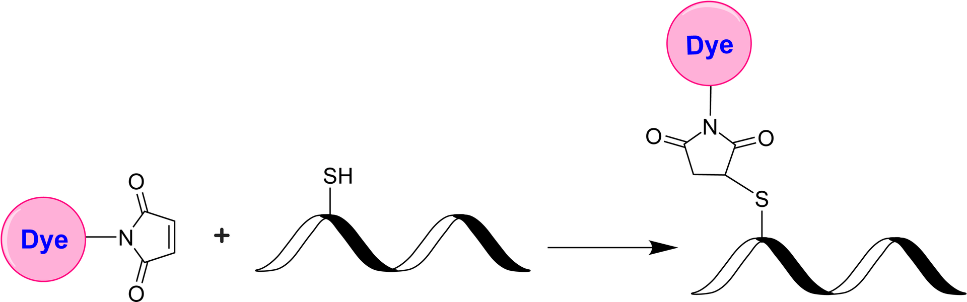
iFluor® 540 maleimide
iFluor® 540 maleimide selectively reacts with thiol group of a biomolecule. It is widely used to label the reduced antibodies and other thiol-containing biomolecules such as peptides and thiol-modified oligos. iFluor®540 is a fluorophore from the iFluor® family, which is known for its bright red fluorescence and compatibility with various fluorescence techniques and instruments. When excited with light in the green to yellow range (around 530 to 545 nm), iFluor® 540 emits red fluorescence with an emission peak typically around 560 to 570 nanometers. iFluor® 540 can be conjugated to a variety of biomolecules, including antibodies, proteins, nucleic acids, and small molecules, enabling their visualization and tracking in cells and tissues. It is commonly used in fluorescence microscopy, immunohistochemistry, flow cytometry, and other fluorescence-based assays.
| Catalog | Size | Price | Quantity |
|---|---|---|---|
| 1427 | 1 mg | Price |

Physical properties
| Molecular weight | 777.82 |
| Solvent | DMSO |
Spectral properties
| Correction factor (260 nm) | 0.26 |
| Correction factor (280 nm) | 0.105 |
| Extinction coefficient (cm -1 M -1) | 80,000 |
| Excitation (nm) | 540 |
| Emission (nm) | 557 |
| Quantum yield | 0.63 |
Storage, safety and handling
| H-phrase | H303, H313, H333 |
| Hazard symbol | XN |
| Intended use | Research Use Only (RUO) |
| R-phrase | R20, R21, R22 |
| Storage | Freeze (< -15 °C); Minimize light exposure |
Contact us
| Telephone | |
| Fax | |
| sales@aatbio.com | |
| International | See distributors |
| Bulk request | Inquire |
| Custom size | Inquire |
| Technical Support | Contact us |
| Request quotation | Request |
| Purchase order | Send to sales@aatbio.com |
| Shipping | Standard overnight for United States, inquire for international |
Page updated on July 14, 2026