logo
AAT Bioquest

iFluor® 665 maleimide

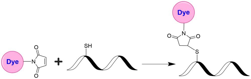
AAT Bioquest's iFluor® dyes are optimized for labeling proteins, particularly antibodies. These dyes are bright, photostable, and have minimal quenching on proteins. They can be well excited by the major laser lines of fluorescence instruments (e.g., 350, 405, 488, 532, 555, 633, and 647 nm). The iFluor® 665 family has spectral properties similar to those of Alexa Fluor® 660 (Alexa Fluor® is the trademark of Invitrogen). In addition, the fluorescence of iFluor® 665 is pH-insensitive over a broad range, pH 3-11. These spectral characteristics make this new dye family an excellent alternative to Alexa Fluor® 660. Under the same conditions, iFluor® 665 gives a stronger fluorescence signal on some antibodies we tested. iFluor® 665 maleimide is reasonably stable and shows good reactivity and selectivity with protein thiol groups even under neutral or slightly acidic conditions.

Example protocol

PREPARATION OF STOCK SOLUTIONS

Unless otherwise noted, all unused stock solutions should be divided into single-use aliquots and stored at -20 °C after preparation. Avoid repeated freeze-thaw cycles.

1. iFluor™ 665 maleimide stock solution (Solution B)
Add anhydrous DMSO into the vial of iFluor™ 665 maleimide to make a 10 mM stock solution. Mix well by pipetting or vortex.
Note     Prepare the dye stock solution (Solution B) before starting the conjugation. Use promptly. Extended storage of the dye stock solution may reduce the dye activity. Solution B can be stored in freezer for upto 4 weeks when kept from light and moisture. Avoid freeze-thaw cycles.


2. Protein stock solution (Solution A)
Mix 100 µL of a reaction buffer (e.g., 100 mM MES buffer with pH ~6.0) with 900 µL of the target protein solution (e.g. antibody, protein concentration >2 mg/mL if possible) to give 1 mL protein labeling stock solution.
Note     The pH of the protein solution (Solution A) should be 6.5 ± 0.5.
Note     Impure antibodies or antibodies stabilized with bovine serum albumin (BSA) or other proteins will not be labeled well.
Note     The conjugation efficiency is significantly reduced if the protein concentration is less than 2 mg/mL. For optimal labeling efficiency the final protein concentration range of 2-10 mg/mL is recommended.

Optional: if your protein does not contain a free cysteine, you must treat your protein with DTT or TCEP to generate a thiol group. DTT or TCEP are used for converting a disulfide bond to two free thiol groups. If DTT is used you must remove free DTT by dialysis or gel filtration before conjugating a dye maleimide to your protein. Following is a sample protocol for generating a free thiol group:
  1. Prepare a fresh solution of 1 M DTT (15.4 mg/100 µL) in distilled water.
  2. Make IgG solution in 20 mM DTT: add 20 µL of DTT stock per ml of IgG solution while mixing. Let stand at room temp for 30 minutes without additional mixing (to minimize reoxidation of cysteines to cystines).
  3. Pass the reduced IgG over a filtration column pre-equilibrated with "Exchange Buffer". Collect 0.25 mL fractions off the column.
  4. Determine the protein concentrations and pool the fractions with the majority of the IgG. This can be done either spectrophotometrically or colorimetrically.
  5. Carry out the conjugation as soon as possible after this step (see Sample Experiment Protocol).
    Note     IgG solutions should be >4 mg/mL for the best results. The antibody should be concentrated if less than 2 mg/mL. Include an extra 10% for losses on the buffer exchange column.
    Note     The reduction can be carried out in almost any buffers from pH 7-7.5, e.g., MES, phosphate or TRIS buffers.
    Note     Steps 3 and 4 can be replaced by dialysis. 

SAMPLE EXPERIMENTAL PROTOCOL

This labeling protocol was developed for the conjugate of Goat anti-mouse IgG with iFluor™ 665 maleimide. You might need further optimization for your particular proteins.
Note     Each protein requires distinct dye/protein ratio, which also depends on the properties of dyes. Over labeling of a protein could detrimentally affects its binding affinity while the protein conjugates of low dye/protein ratio gives reduced sensitivity.


Run conjugation reaction
  1. Use 10:1 molar ratio of Solution B (dye)/Solution A (protein) as the starting point:  Add 5 µL of the dye stock solution (Solution B, assuming the dye stock solution is 10 mM) into the vial of the protein solution (95 µL of Solution A) with effective shaking. The concentration of the protein is ~0.05 mM assuming the protein concentration is 10 mg/mL and the molecular weight of the protein is ~200KD.
    Note     We recommend to use 10:1 molar ratio of Solution B (dye)/Solution A (protein). If it is too less or too high, determine the optimal dye/protein ratio at 5:1, 15:1 and 20:1 respectively.
  2. Continue to rotate or shake the reaction mixture at room temperature for 30-60 minutes. 

Purify the conjugation
The following protocol is an example of dye-protein conjugate purification by using a Sephadex G-25 column.
  1. Prepare Sephadex G-25 column according to the manufacture instruction.
  2. Load the reaction mixture (From "Run conjugation reaction") to the top of the Sephadex G-25 column.
  3. Add PBS (pH 7.2-7.4) as soon as the sample runs just below the top resin surface.
  4. Add more PBS (pH 7.2-7.4) to the desired sample to complete the column purification. Combine the fractions that contain the desired dye-protein conjugate.
    Note     For immediate use, the dye-protein conjugate need be diluted with staining buffer, and aliquoted for multiple uses.
    Note     For longer term storage, dye-protein conjugate solution need be concentrated or freeze dried. 

Spectrum

Product family

NameExcitation (nm)Emission (nm)Extinction coefficient (cm -1 M -1)Quantum yieldCorrection Factor (260 nm)Correction Factor (280 nm)
iFluor® 350 maleimide3454502000010.9510.830.23
iFluor® 488 maleimide4915167500010.910.210.11
iFluor® 555 maleimide55757010000010.6410.230.14
iFluor® 647 maleimide65667025000010.2510.030.03
iFluor® 680 maleimide68470122000010.2310.0970.094
iFluor® 700 maleimide69071322000010.2310.090.04
iFluor® 750 maleimide75777927500010.1210.0440.039
iFluor® 790 maleimide78781225000010.1310.10.09
iFluor® 800 maleimide80182025000010.1110.030.08
iFluor® 810 maleimide81182225000010.0510.090.15
iFluor® 820 maleimide82285025000010.110.16
iFluor® 860 maleimide85387825000010.10.14
iFluor® 532 maleimide5375609000010.6810.260.16
iFluor® 594 maleimide58760320000010.5310.050.04
iFluor® 405 maleimide4034273700010.9110.480.77
iFluor® 430 maleimide4334984000010.7810.680.3
iFluor® 568 maleimide56858710000010.5710.340.15
iFluor® 633 maleimide64065425000010.2910.0620.044
iFluor® 450 maleimide4515024000010.8210.450.27
iFluor® 460 maleimide468493800001~0.810.980.46
iFluor® 546 maleimide54155710000010.6710.250.15
iFluor® 840 maleimide8368792000001-0.20.09
iFluor® 770 maleimide77779725000010.160.090.08
iFluor® 780 maleimide78480825000010.1610.130.12
iFluor® 830 maleimide830867----
iFluor® 514 maleimide5115277500010.8310.2650.116
iFluor® 660 maleimide66367825000010.2610.070.08
iFluor® 670 maleimide67168220000010.5510.030.033
iFluor® 720 maleimide71674024000010.1410.150.13
Show More (20)

References

View all 7 references: Citation Explorer
Recognition of Invasive Prostate Cancer Using a GHRL Polypeptide Probe Targeting GHSR in a Mouse Model In Vivo.
Authors: Ye, Huamao and Yang, Yue and Chen, Rui and Shi, Xiaolei and Fang, Yu and Yang, Jun and Dong, Yuanzhen and Chen, Lili and Xia, Jianghua and Wang, Chao and Yang, Chenghua and Feng, Jun and Wang, Yang and Feng, Xiang and Lü, Chen
Journal: Current pharmaceutical design (2020): 1614-1621
A rapid sensitive, flow cytometry-based method for the detection of Plasmodium vivax-infected blood cells.
Authors: Roobsoong, Wanlapa and Maher, Steven P and Rachaphaew, Nattawan and Barnes, Samantha J and Williamson, Kim C and Sattabongkot, Jetsumon and Adams, John H
Journal: Malaria journal (2014): 55
Reengineering the optical absorption cross-section of photosynthetic reaction centers.
Authors: Dutta, Palash K and Lin, Su and Loskutov, Andrey and Levenberg, Symon and Jun, Daniel and Saer, Rafael and Beatty, J Thomas and Liu, Yan and Yan, Hao and Woodbury, Neal W
Journal: Journal of the American Chemical Society (2014): 4599-604
Comparison of a chimeric anti-carcinoembryonic antigen antibody conjugated with visible or near-infrared fluorescent dyes for imaging pancreatic cancer in orthotopic nude mouse models.
Authors: Maawy, Ali A and Hiroshima, Yukihiko and Kaushal, Sharmeela and Luiken, George A and Hoffman, Robert M and Bouvet, Michael
Journal: Journal of biomedical optics (2013): 126016
Nucleic acid sandwich hybridization assay with quantum dot-induced fluorescence resonance energy transfer for pathogen detection.
Authors: Chou, Cheng-Chung and Huang, Yi-Han
Journal: Sensors (Basel, Switzerland) (2012): 16660-72
Page updated on September 19, 2024

Ordering information

Price
Unit size
Catalog Number1554
Quantity
Add to cart

Additional ordering information

Telephone1-800-990-8053
Fax1-800-609-2943
Emailsales@aatbio.com
InternationalSee distributors
Bulk requestInquire
Custom sizeInquire
Technical SupportContact us
Purchase orderSend to sales@aatbio.com
ShippingStandard overnight for United States, inquire for international
Request quotation

Physical properties

Molecular weight

1147.22

Solvent

DMSO

Spectral properties

Absorbance (nm)

661

Correction Factor (260 nm)

0.12

Correction Factor (280 nm)

0.09

Extinction coefficient (cm -1 M -1)

110,0001

Excitation (nm)

667

Emission (nm)

692

Quantum yield

0.221

Storage, safety and handling

H-phraseH303, H313, H333
Hazard symbolXN
Intended useResearch Use Only (RUO)
R-phraseR20, R21, R22

Storage

Freeze (< -15 °C); Minimize light exposure
UNSPSC12171501
Fluorescent dye maleimides are the most popular tool for conjugating dyes to a peptide, protein, antibody, thiol-modified oligonucleotide, or nucleic acid through their SH group. Maleimides react readily with the thiol group of proteins, thiol-modified oligonucleotides, and other thiol-containing molecules under neutral conditions. The resulting dye conjugates are quite stable.