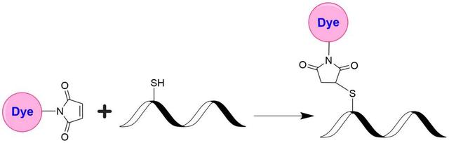
iFluor® 780 maleimide
Example protocol
PREPARATION OF STOCK SOLUTIONS
Unless otherwise noted, all unused stock solutions should be divided into single-use aliquots and stored at -20 °C after preparation. Avoid repeated freeze-thaw cycles
- Allow the vial of iFluor Dye maleimide to warm up to room temperature.
- Add anhydrous DMSO to the vial to prepare a 10 mM dye stock solution.
- Vortex the vial briefly to fully dissolve the dye, and then centrifuge to collect the dye at the bottom of the vial.
- Protect all stock solutions from light as much as possible by wrapping containers in aluminum foil.
- If your protein already contains a thiol group, prepare the protein at 50-100 uM (for example: 5mg/ml BSA is ~75uM) in 50~100 mM MES buffer or buffers of your choice with pH 6.5~7.0.
- If labeling with an intact antibody, reduction of disulfide bonds need to be carried out before maleimide reaction. Prepare antibody in 2-10 mg/ml in a suitable buffer with pH 7.0–7.5. A 10-fold molar excess of a reducing agent such as DTT or TCEP is added to the antibody. If DTT is used, it must be removed by dialysis or desalting to a suitable buffer with pH 6.5~7.0 prior to conjugation. If TCEP is used, it is not necessary to remove excess TCEP during conjugation with maleimides, however, removal of TCEP by dialysis or desalting prior to conjugation gives the better labeling efficiency.
Below is a sample protocol for generating free thiol groups on antibody:-
Prepare 2-10mg/ml IgG solution in PBS.
-
Prepare a fresh solution of 1 M DTT (15.4 mg/100 µL) in distilled water.
-
Add 1- 20 µL of DTT stock per ml of IgG solution while mixing.
-
Let the solution stand at room temperature for 30 minutes without additional mixing (to minimize the re-oxidation of cysteines to cystines).
-
Pass the reduced IgG over a filtration column pre-equilibrated with 50 mM MES buffer (pH=6.5) to remove excess DTT.
-
Determine the antibody concentrations. This can be done either spectrophotometrically or colorimetrically.
-
Carry out the conjugation as soon as possible after this step.
Note: For the best results, IgG solutions should be > 2 mg/mL.
Note: The reduction can be carried out in almost any buffer from pH 7 to 7.5, e.g., MES, phosphate, or TRIS buffers.Note: Steps 5 can be replaced by dialysis.
-
- If your protein doesn’t have a free thiol group or disulfide bond to reduce, a thiolation modification need to be carried out before maleimide conjugation (for example: using 2-Iminothiolane or 2-IT) to introduce sulfhydryl (-SH) groups to the original amino groups on protein.
SAMPLE EXPERIMENTAL PROTOCOL
This labeling protocol was developed for the labeling IgG with iFluor® Dye maleimide. Further optimization may be required for your specific proteins.
Note: Each protein requires a distinct dye/protein ratio, which also depends on the properties of dyes. Over-labeling of a protein could detrimentally affect its binding affinity while the protein conjugates of low dye/protein ratio give reduced sensitivity.
- Use a 10~20:1 molar ratio of iFluor Dye Maleimide dye: IgG as the starting point. While stirring or vortexing the protein solution, add a volume of dye stock solution to result in a dye: protein molar ratio of 10-20. For example, for 5mg/ml IgG (~33 uM), you would add dye to a final concentration of 0.33-0.66 mM.
Note: We recommend using a 10:1 molar ratio of dye to protein. If the ratio is too low or too high, determine the optimal dye/protein ratio at 5:1, 15:1, and 20:1, respectively. - Continue to rotate or shake the reaction mixture at room temperature for 30-60 minutes.
Purify the conjugate on a gel filtration column, such as a Sephadex G-25 column or equivalent matrix, or by extensive dialysis at 4°C in an appropriate buffer.
Recommended AAT Desalting Columns:
| Volume of Reaction | Catalog# |
| 0.6-1.0mL | Cat#60504: PD-10 Column https://www.aatbio.com/products/readiuse-disposable-pd-10-desalting-column?unit=60504 |
| ~0.1mL | Cat#60500: Spin Column https://www.aatbio.com/products/readiuse-bio-gel-p-6-spin-column?unit=60500 |
Determining the Degree of Substitution (DOS) is crucial in characterizing dye-labeled proteins. Lower DOS proteins tend to have weaker fluorescence, but higher DOS proteins may also have reduced fluorescence. For most antibodies, the optimal DOS is between 2 and 10, depending on the dye and protein properties. For effective labeling, the degree of substitution should be controlled to have 5-8 moles of iFluor® 780 maleimide to one mole of antibody. The following steps are used to determine the DOS of iFluor® 780 maleimide-labeled proteins:
- Measure absorption— To measure the absorption spectrum of a dye-protein conjugate, the sample concentration should be kept between 1 and 10 µM (For example: IgG conjugate: 10uM is ~1.5mg/ml), depending on the dye's extinction coefficient.
- Read OD (absorbance) at 280 nm and dye maximum absorption (ƛ max = 784 nm for iFluor® 780 dyes). For most spectrophotometers, the sample (from the column fractions) must be diluted with de-ionized water so that the OD values range from 0.1 to 0.9. The O.D. (absorbance) at 280 nm is the maximum absorption of protein, while 784 nm is the maximum absorption of iFluor® 780 maleimide. To obtain accurate DOS, ensure the conjugate is free of the non-conjugated dye.
- Calculate DOS using our DOS calculator: https://www.aatbio.com/tools/degree-of-labeling-calculator
Calculators
Common stock solution preparation
| 0.1 mg | 0.5 mg | 1 mg | 5 mg | 10 mg | |
| 1 mM | 69.451 µL | 347.256 µL | 694.512 µL | 3.473 mL | 6.945 mL |
| 5 mM | 13.89 µL | 69.451 µL | 138.902 µL | 694.512 µL | 1.389 mL |
| 10 mM | 6.945 µL | 34.726 µL | 69.451 µL | 347.256 µL | 694.512 µL |
Molarity calculator
| Mass (Calculate) | Molecular weight | Volume (Calculate) | Concentration (Calculate) | Moles | ||||
| / | = | x | = |
Spectrum

Product family
| Name | Excitation (nm) | Emission (nm) | Extinction coefficient (cm -1 M -1) | Quantum yield | Correction Factor (260 nm) | Correction Factor (280 nm) |
| iFluor® 350 maleimide | 345 | 450 | 200001 | 0.951 | 0.83 | 0.23 |
| iFluor® 405 maleimide | 403 | 427 | 370001 | 0.911 | 0.48 | 0.77 |
| iFluor® 430 maleimide | 433 | 498 | 400001 | 0.781 | 0.68 | 0.3 |
| iFluor® 450 maleimide | 451 | 502 | 400001 | 0.821 | 0.45 | 0.27 |
| iFluor® 460 maleimide | 468 | 493 | 800001 | ~0.81 | 0.98 | 0.46 |
| iFluor® 488 maleimide | 491 | 516 | 750001 | 0.91 | 0.21 | 0.11 |
| iFluor® 510 maleimide | 511 | 530 | - | - | - | - |
| iFluor® 514 maleimide | 511 | 527 | 750001 | 0.831 | 0.265 | 0.116 |
| iFluor® 532 maleimide | 537 | 560 | 900001 | 0.681 | 0.26 | 0.16 |
Show More (25) | ||||||
References
Authors: Morlandt, Anthony B and Moore, Lindsay S and Johnson, Aubrey O and Smith, Caris M and Stevens, Todd M and Warram, Jason M and MacDougall, Mary and Rosenthal, Eben L and Amm, Hope M
Journal: Journal of oral and maxillofacial surgery : official journal of the American Association of Oral and Maxillofacial Surgeons (2020): 1736-1747
Authors: de Oliveira, Cristiane and Patel, Krutika and Mishra, Vivek and Trivedi, Ram N and Noel, Pawan and Singh, Abhilasha and Yaron, Jordan R and Singh, Vijay P
Journal: PloS one (2016): e0149073
Authors: Conner, Kip P and Rock, Brooke M and Kwon, Gayle K and Balthasar, Joseph P and Abuqayyas, Lubna and Wienkers, Larry C and Rock, Dan A
Journal: Drug metabolism and disposition: the biological fate of chemicals (2014): 1906-13
Authors: Bhattacharyya, Sibaprasad and Patel, Nimit and Wei, Ling and Riffle, Lisa A and Kalen, Joseph D and Hill, G Craig and Jacobs, Paula M and Zinn, Kurt R and Rosenthal, Eben
Journal: MedChemComm (2014): 1337-1346
Authors: Ricci, Ugo and Sani, Ilaria and Klintschar, Michael and Cerri, Nicoletta and De Ferrari, Francesco and Giovannucci Uzielli, Maria Luisa
Journal: Croatian medical journal (2003): 299-305